Determining calcium content from spectral ct data

a computed tomography and calcium content technology, applied in image analysis, image enhancement, healthcare informatics, etc., can solve the problems of residual breathing or cardiac motion, myocardial infarction, and deterioration of calcium scores derived from such volumetric images acquired by a known technique, and achieve good, efficient and/or robust methods

Active Publication Date: 2019-09-26
KONINKLJIJKE PHILIPS NV
View PDF0 Cites 3 Cited by
  • Summary
  • Abstract
  • Description
  • Claims
  • Application Information

AI Technical Summary

Benefits of technology

[0008]It is an object of embodiments of the present invention to provide good, efficient and / or robust methods and means for determining, e.g. calculating and / or estimating, a calcium content from CT data.

Problems solved by technology

When an atherosclerosis condition is allowed to develop further, accumulated material adhering to the walls may release suddenly to form intraluminal thrombi which can occlude a coronary artery supplying the heart muscle, thus causing a myocardial infarction, or which can form a thromboembolism.
Furthermore, residual breathing or cardiac motion may deteriorate the calcium scores derived from such volumetric images acquired by a known technique, e.g. due to image blurring.

Method used


Image

  • Determining calcium content from spectral ct data
  • Determining calcium content from spectral ct data

Examples


Embodiment Construction

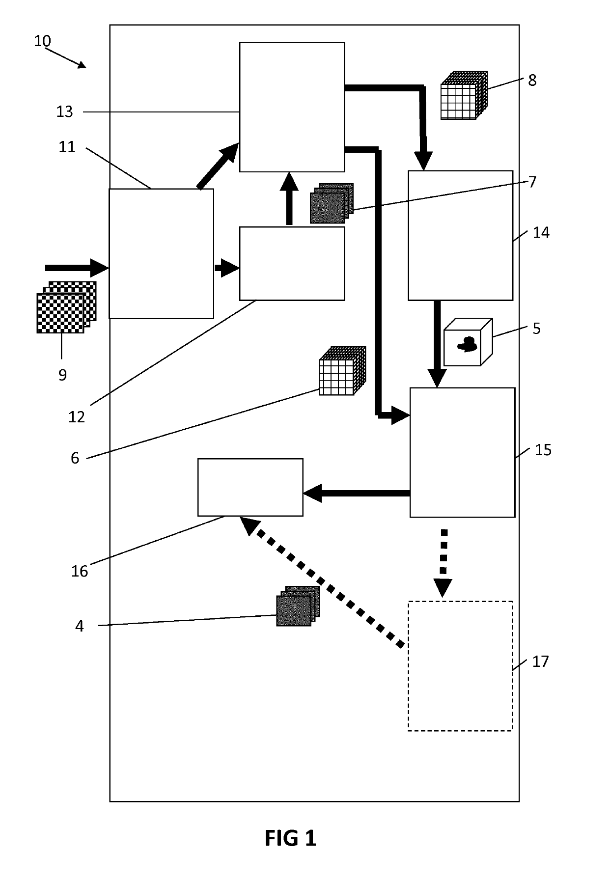
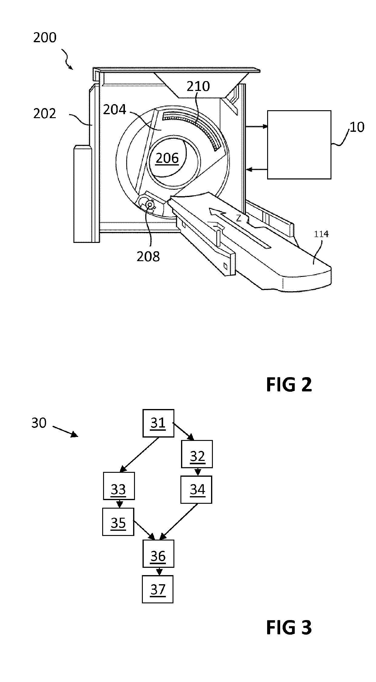
[0041]The present invention will be described with respect to particular embodiments and with reference to certain drawings but the invention is not limited thereto but only by the claims. The drawings described are only schematic and are non-limiting. In the drawings, the size of some of the elements may be exaggerated and not drawn on scale for illustrative purposes. The dimensions and the relative dimensions do not correspond to actual reductions to practice of the invention.

[0042]Furthermore, the terms first, second and the like in the description and in the claims, are used for distinguishing between similar elements and not necessarily for describing a sequence, either temporally, spatially, in ranking or in any other manner. It is to be understood that the terms so used are interchangeable under appropriate circumstances and that the embodiments of the invention described herein are capable of operation in other sequences than described or illustrated herein.

[0043]Moreover, t...


PUM


Abstract

Present invention relates to devices and methods for determining a calcium content by analyzing cardiac spectral CT data. CT projection data (9), obtainable by scanning a cardiac region of a subject using a spectral CT scanning unit, is modelled (12) by applying a material decomposition algorithm to the projection data to provide a calcium-specific component. Tomographic reconstructions (13) of the projection data, to provide a first 3D image (8), and of the calcium-specific component, to provide a second 3D image (6), are performed. The first 3D image (8) is segmented (14) to provide an image mask (5) corresponding to a cardiovascular structure of interest, a part of the second 3D image (6) is selected (15) based on the image mask (5), and a calcium content is calculated (16) in the cardiovascular structure of interest based on the selected part of the second 3D image (6).

Description

FIELD OF THE INVENTION[0001]The invention relates to the field of computed tomography (CT) imaging and related image processing. More specifically it relates to a system and a method for analyzing images obtained by computed tomography, and a computer program product for implementing such method.BACKGROUND OF THE INVENTION[0002]In radiologic cardiovascular examination, determining a calcium content may be particularly useful, e.g. for determining a coronary artery calcium score. Such calcium score is a measurement known in the art for determining, e.g. estimating and / or quantifying, the amount of calcium that is present in the walls of arteries that supply the heart muscle. This amount of calcium is correlated with hardening of the arterial wall, e.g. may indicate an arteriosclerotic vascular disease, such as, particularly, atherosclerosis. In atherosclerosis, a plaque is formed on the arterial wall, which may comprise a nodular accumulation of atheroma, crystal deposits of choleste...

Claims


Application Information

Patent Timeline
Patent Type & Authority Applications(United States)
IPC IPC(8): G06T7/00G16H30/40G06T11/00
CPCG06T2211/404G06T2207/30048G16H30/40G06T2211/408G06T2207/30101G06T2207/10081G06T11/005G06T7/0012A61B5/02007
Inventor SCHAEFER, DIRKDAERR, HEINERKOEHLER, THOMASROESSL, EWALD
Owner KONINKLJIJKE PHILIPS NV
Why Eureka
Try Eureka