Electron Microscope and Specimen Tilt Angle Adjustment Method

a technology of electric microscope and specimen, which is applied in the direction of basic electric elements, electric discharge tubes, electrical equipment, etc., can solve the problems of automatic adjustment of specimen tilt angle, measurement error, and inability to adjust specimen tilt angle for the first time, so as to achieve the effect of easily adjusting the specimen tilt angl

Active Publication Date: 2019-04-18
JEOL LTD
View PDF3 Cites 5 Cited by
  • Summary
  • Abstract
  • Description
  • Claims
  • Application Information

AI Technical Summary

Benefits of technology

[0011]The invention provides an electron microscope and a specimen tilt angle adjustment method capable of adjusting a specimen tilt angle easily.

Problems solved by technology

This is because a very small difference between a tilt angle of a specimen and an incidence direction of an electron beam may cause a measurement error.
This adjustment can be realized in several seconds by skilled operators, whereas the adjustment may require a considerable time for inexperienced operators.
Therefore, it is not possible to adjust the specimen tilt angle automatically if a material is not registered in the computer, and thus, the method has a low versatility problem.
Moreover, an operator has to register information on a material type and a crystal orientation into the computer whenever adjustment is performed, and thus, the method has a low operability problem.
Due to this, it requires a considerable time to tilt the specimen, and thus, the adjustment of the specimen tilt angle takes time.

Method used


Image

  • Electron Microscope and Specimen Tilt Angle Adjustment Method
  • Electron Microscope and Specimen Tilt Angle Adjustment Method
  • Electron Microscope and Specimen Tilt Angle Adjustment Method

Examples


first modification

5.1. First Modification

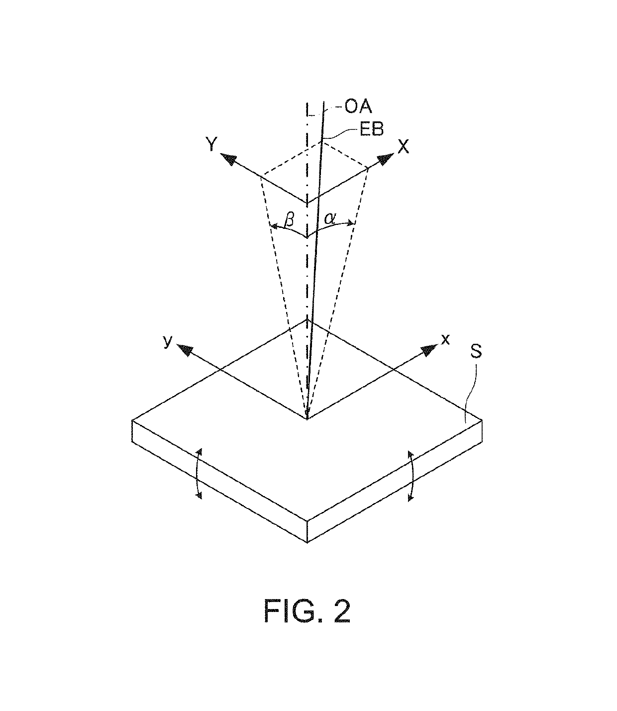
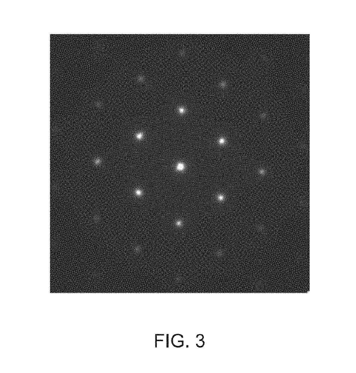
[0134]First, a first modification will be described. The specimen tilt angle adjustment method according to the above-described embodiments calculates an electron diffraction spot coordinate of each of a plurality of acquired electron diffraction patterns, calculates the radius of an approximate circle by performing circular approximation on the electron diffraction pattern on the basis of the electron diffraction spot coordinate, calculates an incidence angle at which the calculated radius of the approximate circle is minimized by fitting, and calculates the tilt angle from the calculated incidence angle.

[0135]In contrast, in the first modification, an electron diffraction spot coordinate is calculated for each of a plurality of acquired electron diffraction patterns, the central coordinate of an approximate circle is calculated by performing circular approximation on the electron diffraction pattern on the basis of the electron diffraction spot coordinate, t...

second modification

5.2. Second Modification

[0147]Next, a second modification will be described.

[0148]In the above-described embodiments, when changing the specimen tilt angle to the optimal tilt angles STx and STy, a focus shift resulting from movement of a field of view and movement of a specimen height associated with tilting of the specimen may occur. Therefore, an amorphous material portion may be irradiated with an electron beam or a different adjacent crystalline material may be irradiated with an electron beam due to tilting of the specimen.

[0149]Therefore, in the controller 34, information on a movement amount (two-dimensional information) of an observation field of view of the specimen S due to tilting of the specimen is stored in the storage unit 346 as correction data for respective specimen tilting axes. The controller 34 (the control unit 340b) reads the correction data from the storage unit 346 whenever changing the specimen tilt angle to calculate a correction value by interpolation fro...


PUM


Abstract

An electron microscope includes: an irradiation lens system that irradiates a specimen with an electron beam; an irradiation system deflector that deflects an electron beam incident on the specimen; a specimen tilting mechanism that tilts the specimen; an imaging lens system that forms an electron diffraction pattern or an electron microscope image by using an electron having passed through the specimen; an imaging device that acquires the electron diffraction pattern or the electron microscope image formed by the imaging lens system; and a controller that controls the irradiation system deflector and the specimen tilting mechanism. The controller performs: a process of acquiring a plurality of electron diffraction patters formed by using electron beams having different incidence angles to the specimen, the different incidence angles having been obtained by deflecting the electron beams incident on the specimen by using the irradiation system deflector; a process of calculating a tilt angle of the specimen based on the plurality of electron diffraction patterns; and a process of controlling the specimen tilting mechanism so that the specimen has the calculated tilt angle.

Description

[0001]Japanese Patent Application No. 2017-200803, filed on Oct. 17, 2017, is hereby incorporated by reference in its entirety.BACKGROUND OF THE INVENTION[0002]The present invention relates to an electron microscope and a specimen tilt angle adjustment method.[0003]An electron microscope can irradiate a specimen with an electron beam and magnify and image an electron having passed through the specimen with the aid of an imaging electron optical system. In this way, it is possible to observe the specimen with high resolution. Moreover, the electron microscope can examine arrangements of molecules and atoms of a material of a crystalline specimen on the basis of electron diffraction patterns. Furthermore, the electron microscope can scan an electron beam converged narrowly onto a specimen, detect an electron having passed through the specimen from a specific capturing angle, and synchronize a scanning signal with an electron detection signal. In this way, it is possible to acquire a s...

Claims


Application Information

Patent Timeline
Patent Type & Authority Applications(United States)
IPC IPC(8): H01J37/26H01J37/295H01J37/153H01J37/147H01J37/10
CPCH01J37/261H01J37/295H01J37/153H01J37/1471H01J37/10H01J37/265H01J2237/20207H01J2237/221H01J2237/24578H01J2237/2802H01J2237/2805H01J2237/2817H01J2237/2826
Inventor KAWAI, SHUJI
Owner JEOL LTD
Why Eureka
Try Eureka