Model-based segmentation of an anatomical structure

a model-based segmentation and anatomical structure technology, applied in the field of model-based segmentation of anatomical structure, can solve the problems of inadequacies in the fitting of achieve the effects of enhancing the visibility of otherwise poorly visible parts of the anatomical structure, enhancing the visibility of a surface, and enhancing the visibility of said surfa

Active Publication Date: 2017-07-27
KONINKLJIJKE PHILIPS NV
View PDF1 Cites 4 Cited by
  • Summary
  • Abstract
  • Description
  • Claims
  • Application Information

AI Technical Summary

Benefits of technology

[0038]Optionally, the processor is further configured for performing an image enhancement prior to locating the image feature in the image data. By performing an image enhancement, the visibility of otherwise poorly visible parts of the anatomical structure may be enhanced. For example, the image enhancement may be adapted to the type of image feature, the geometric relation to the first part of the deformable model, etc.
[0039]Optionally, the processor is configured for performing a di...

Problems solved by technology

A problem of the known deformable models and ways of applying such models to an anatomical structure in a medical ...

Method used


Image

  • Model-based segmentation of an anatomical structure
  • Model-based segmentation of an anatomical structure
  • Model-based segmentation of an anatomical structure

Examples


Embodiment Construction

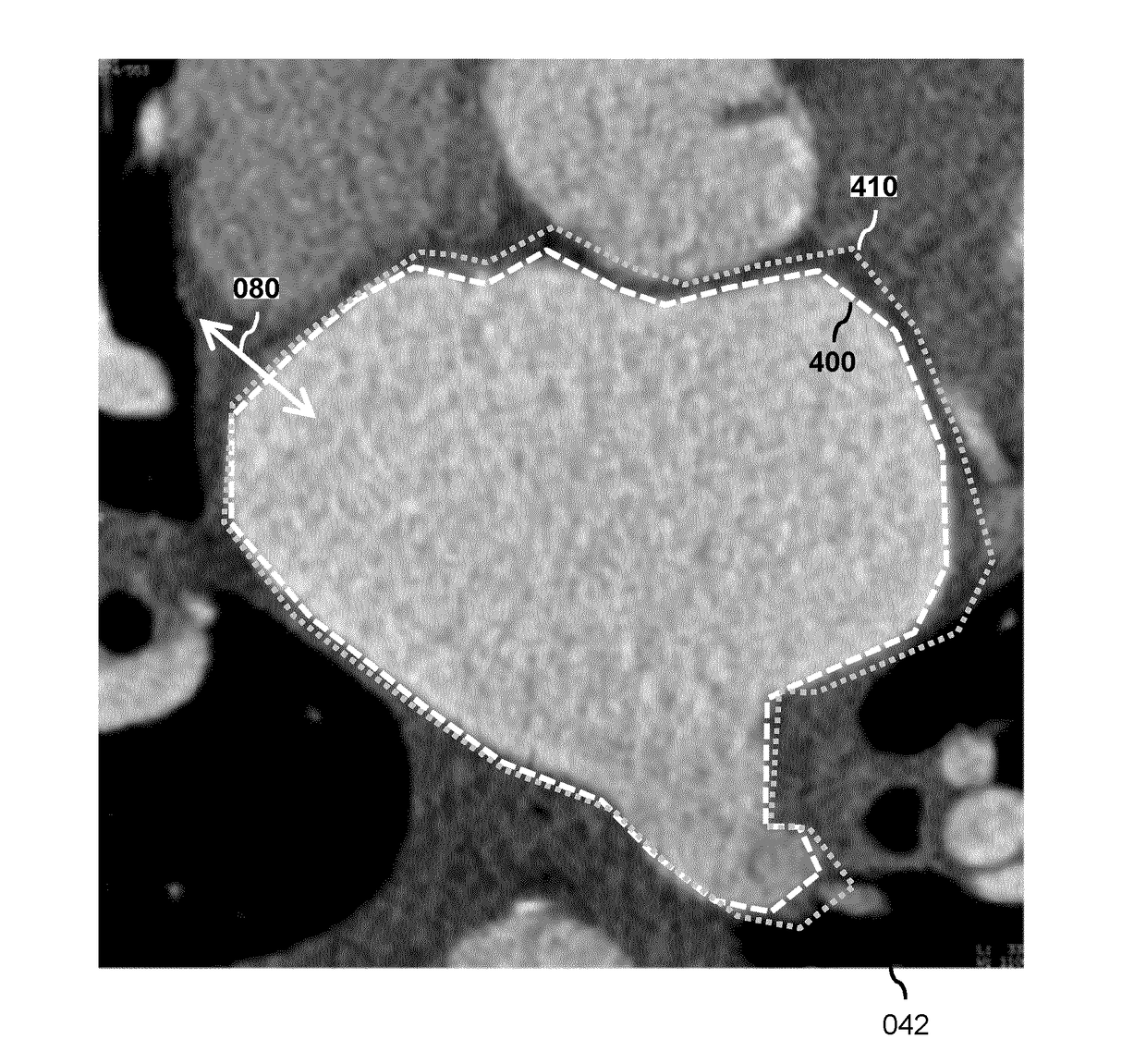
[0064]FIG. 1 shows a system 100 for performing a model-based segmentation of an anatomical structure in a medical image. The system 100 comprises an input 120 for obtaining image data 042 of the medical image. For that purpose, the system 100 is shown to be connected to a database 040. For example, the database 040 may be constituted or part of a Picture Archiving and Communication System (PACS) of a Hospital Information System (HIS) to which the system 100 may be connected or comprised in. The system 100 further comprises a data storage 200 which comprises model data 210 defining a deformable model for segmenting a type of anatomical structure, with the deformable model comprising parts to be fitted to corresponding parts of the anatomical structure. The data storage 200 is shown to be an internal component of the system 100, and may be constituted by, e.g., a disk-based data storage such as a hard disk, a semiconductor-based data storage such as a ROM or RAM memory, a removable st...


PUM


Abstract

A system (100) is provided for performing a model-based segmentation of an anatomical structure in a medical image. The system comprises a processor (140) configured for performing a model-based segmentation of the anatomical structure by applying a deformable model to image data (042). Moreover, definition data (220) is provided which defines a geometric relation between a first part and a second part of the deformable model of which a corresponding first part of the anatomical structure is presumed to be better visible in the image data than a corresponding second part of the anatomical structure. The definition data is then used to adjust a fit of the second part of the deformable model. As a result, a better fit of the second part of the deformable model to the second part of the anatomical structure is obtained despite said part being relatively poorly visible in the image data.

Description

FIELD OF THE INVENTION[0001]The invention relates to a system and a method for performing a model-based segmentation of an anatomical structure in a medical image. The invention further relates to a data storage comprising model data defining a deformable model, to a workstation and imaging apparatus comprising the system and to a computer program product comprising instructions for causing a processor system to perform the method.BACKGROUND OF THE INVENTION[0002]Robust automatic segmentation of various anatomical structures in a medical image is a key enabler in improving clinical workflows. Here, the term segmentation refers to the identification of the anatomical structure in the medical image by, e.g., delineation of the boundaries of the anatomical structure, or by labeling of the voxels enclosed by the boundaries. Once such segmentation has been performed, it is possible to extract clinical parameters such as, in case of a cardiac structure, ventricular mass, ejection fraction...

Claims


Application Information

Patent Timeline
IPC IPC(8): G06T7/00G06T7/12
CPCG06T7/0012G06T2207/30048G06T7/12G06T7/149
Inventor GROTH, ALEXANDRAPETERS, JOCHENWEESE, JUERGENSAALBACH, AXEL
Owner KONINKLJIJKE PHILIPS NV
Why Eureka
Try Eureka