Ultrasound image displaying apparatus and method for displaying ultrasound image

a technology of ultrasound image and display apparatus, applied in the field of ultrasound image, can solve the problems of difficult to grasp the image quality of vascular structure, noise called motion artifacts,

Inactive Publication Date: 2015-12-31
GE MEDICAL SYST GLOBAL TECH CO LLC
View PDF5 Cites 7 Cited by
  • Summary
  • Abstract
  • Description
  • Claims
  • Application Information

AI Technical Summary

Benefits of technology

[0008]According to embodiments of the present invention, the composite data combined using the data of some or all frames in each of which the index related to the amount of the motion artifacts satisfies the prescribed reference is obtained, thus making it possible to display an ultrasound image of desired image quality in terms of the motion artifacts.

Problems solved by technology

Meanwhile in an ultrasound image, noise called motion artifacts may occur due to the pulsation of blood vessels, body motion and the like.
If such noise is included in combined data even one frame, an ultrasound image displayed based on the composite data leads to an image of image quality difficult to grasp a vascular structure.

Method used


Image

  • Ultrasound image displaying apparatus and method for displaying ultrasound image
  • Ultrasound image displaying apparatus and method for displaying ultrasound image
  • Ultrasound image displaying apparatus and method for displaying ultrasound image

Examples


Embodiment Construction

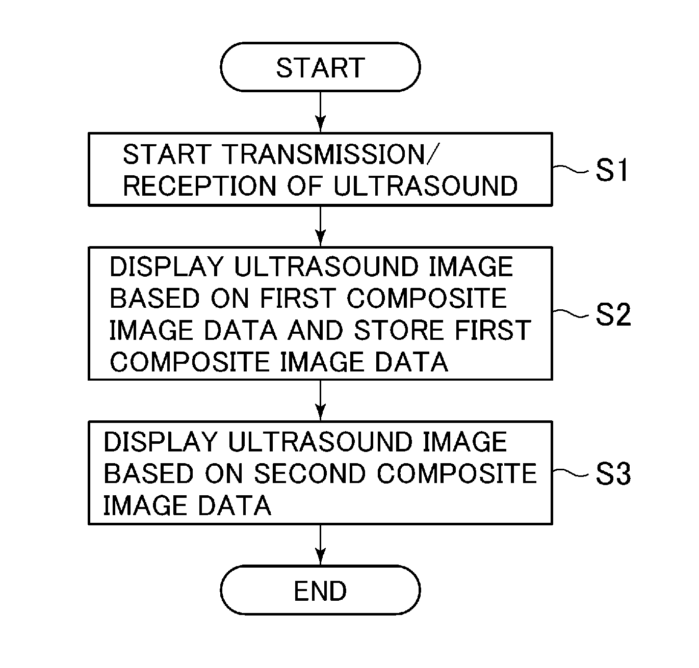
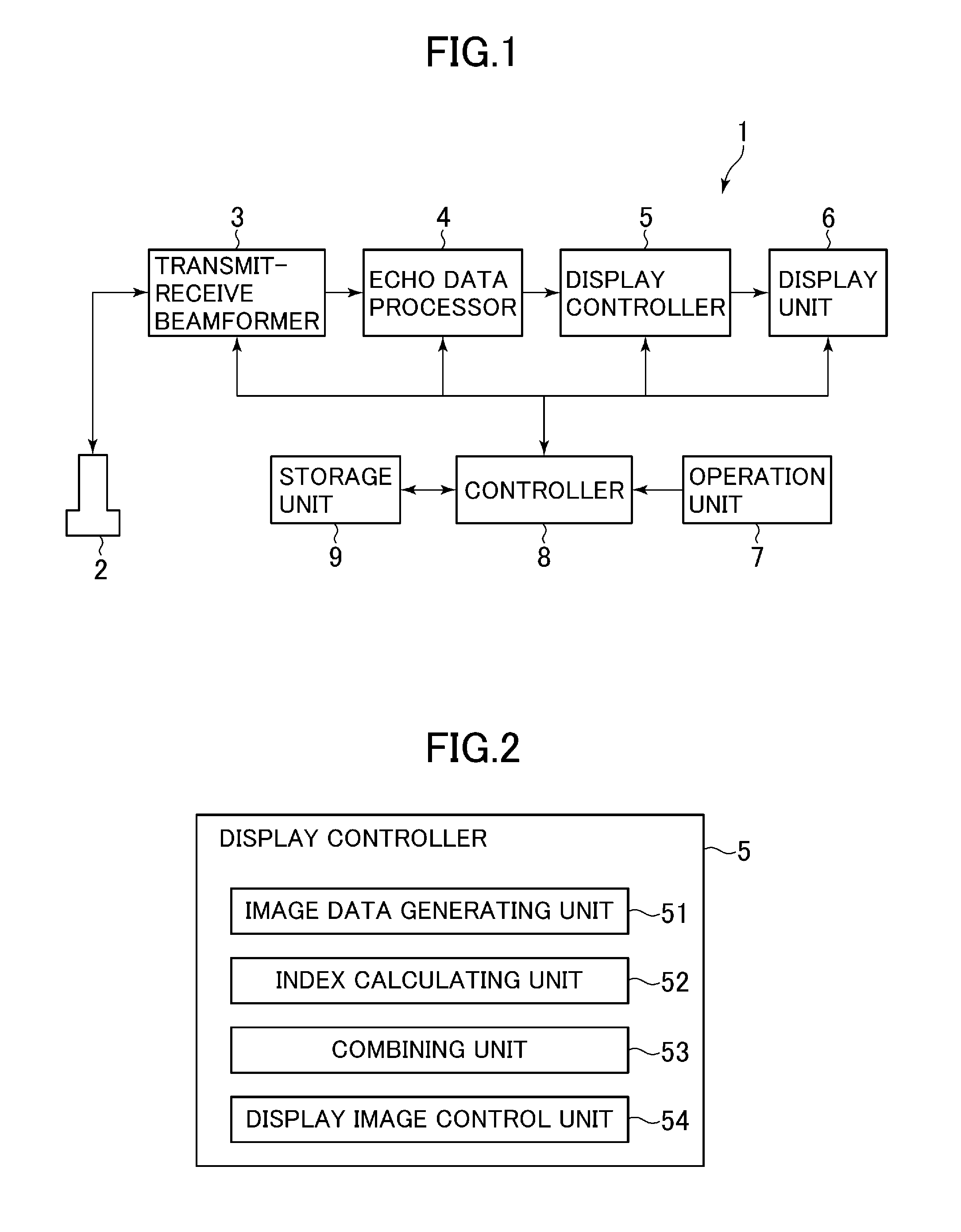
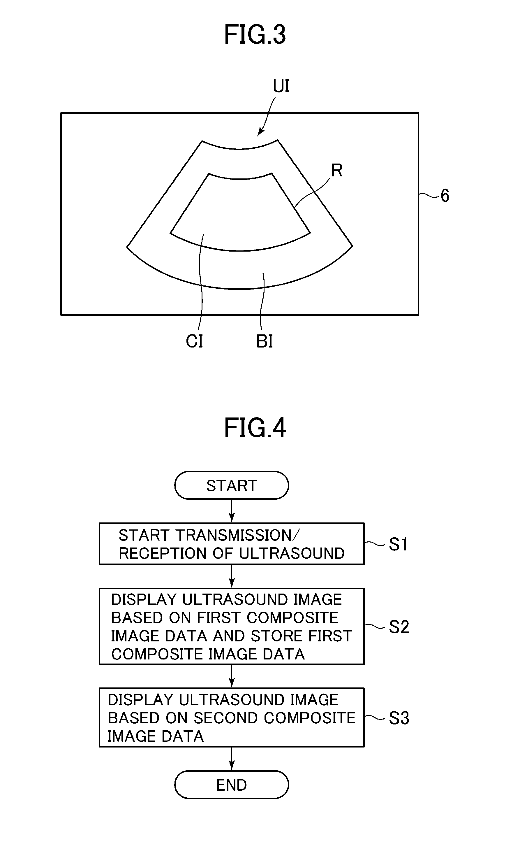
[0018]An embodiment of the invention will hereinafter be described in detail based on FIGS. 1 through 7. An ultrasound diagnostic apparatus 1 shown in FIG. 1 is equipped with an ultrasound probe 2, a transmit-receive beamformer 3, an echo data processor 4, a display controller 5, a display unit 6, an operation unit 7, a controller 8 and a storage unit 9. The ultrasound diagnostic apparatus 1 displays an ultrasound image on the display unit 6. Therefore, the ultrasound diagnostic apparatus includes an ultrasound image displaying apparatus.

[0019]The ultrasound probe 2 is comprised of a plurality of ultrasound transducers (not shown) arranged in array form. The ultrasound probe 2 transmits ultrasound to a subject through the ultrasound transducers and receives its echo signals therein.

[0020]The transmit-receive beamformer 3 supplies an electric signal for transmitting ultrasound from the ultrasound probe 2 under a predetermined scan condition to the ultrasound probe 2, based on a contr...


PUM


Abstract

An ultrasound image displaying apparatus includes: a composite data generating unit which generates composite data in which data based on echo signals obtained by transmitting ultrasound to a subject are multiple-frame combined; a display unit on which an ultrasound image based on the composite data is displayed; and an index calculating unit which calculates an index related to the amount of motion artifacts in the ultrasound image for each frame, based on the data. The composite data generating unit generates composite data combined using data of some or all frames in each of which the index satisfies a prescribed reference.

Description

TECHNICAL FIELD[0001]Embodiments of the present invention relate to an ultrasound image displaying apparatus which generates composite data in which data based on echo signals obtained by transmitting ultrasound to a subject are combined over multiple frames, and a method for displaying an ultrasound image thereof.BACKGROUND ART[0002]For example, when one intends to observe a blood flow in an ultrasound image, the blood flow may be extracted only in a limited way within an ultrasound image of one frame, depending on the position of a transmission / reception surface of ultrasound and imaging timing. There has therefore been disclosed in, for example, a Patent Document 1, an ultrasound diagnostic apparatus which generates composite data in which data based on echo signals are multiple-frame combined, and displays an ultrasound image based on the composite data.[0003]The composite data is comprised of, for example, data indicative of the maximum brightness, of data of plural frames. The...

Claims


Application Information

Patent Timeline
Patent Type & Authority Applications(United States)
IPC IPC(8): G06T5/50G06T7/00G06T7/20
CPCG06T5/50G06T2207/10132G01S15/8979G01S7/52065G01S7/52077A61B8/14A61B8/488A61B8/5276G06T7/0012G06T7/20G06T2207/30104
Inventor KAMIYAMA, NAOHISAHASHIMOTO, HIROSHI
Owner GE MEDICAL SYST GLOBAL TECH CO LLC
Why Eureka
Try Eureka