Methods for isolation of biomarkers from vesicles
a biomarker and vesicles technology, applied in the field of methods for isolation of biomarkers from vesicles, can solve problems such as readingouts that lack the requisite sensitivity
- Summary
- Abstract
- Description
- Claims
- Application Information
AI Technical Summary
Problems solved by technology
Method used
Image
Examples
example 1
of Filters for the Capacity of Exosome Collection
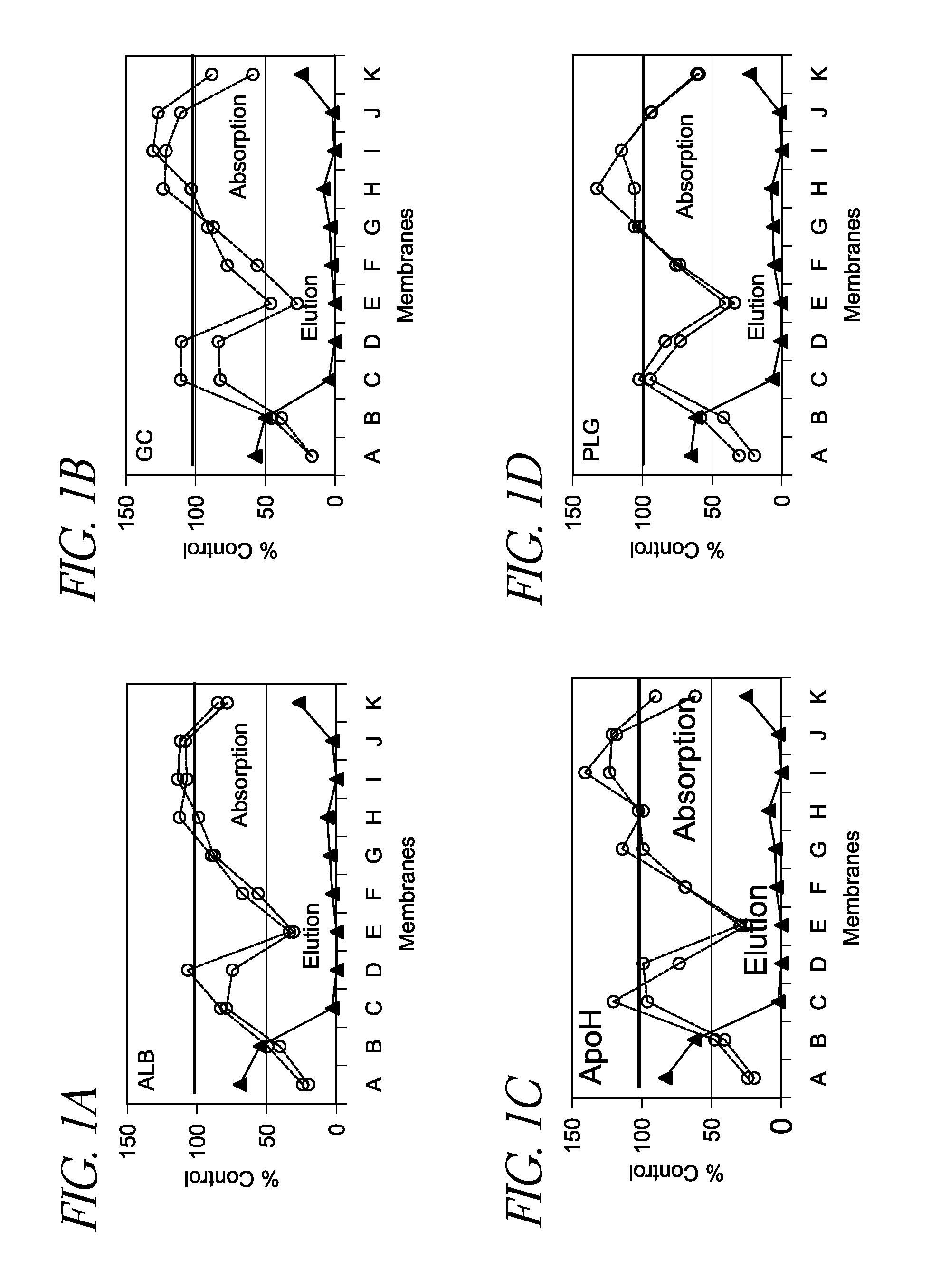
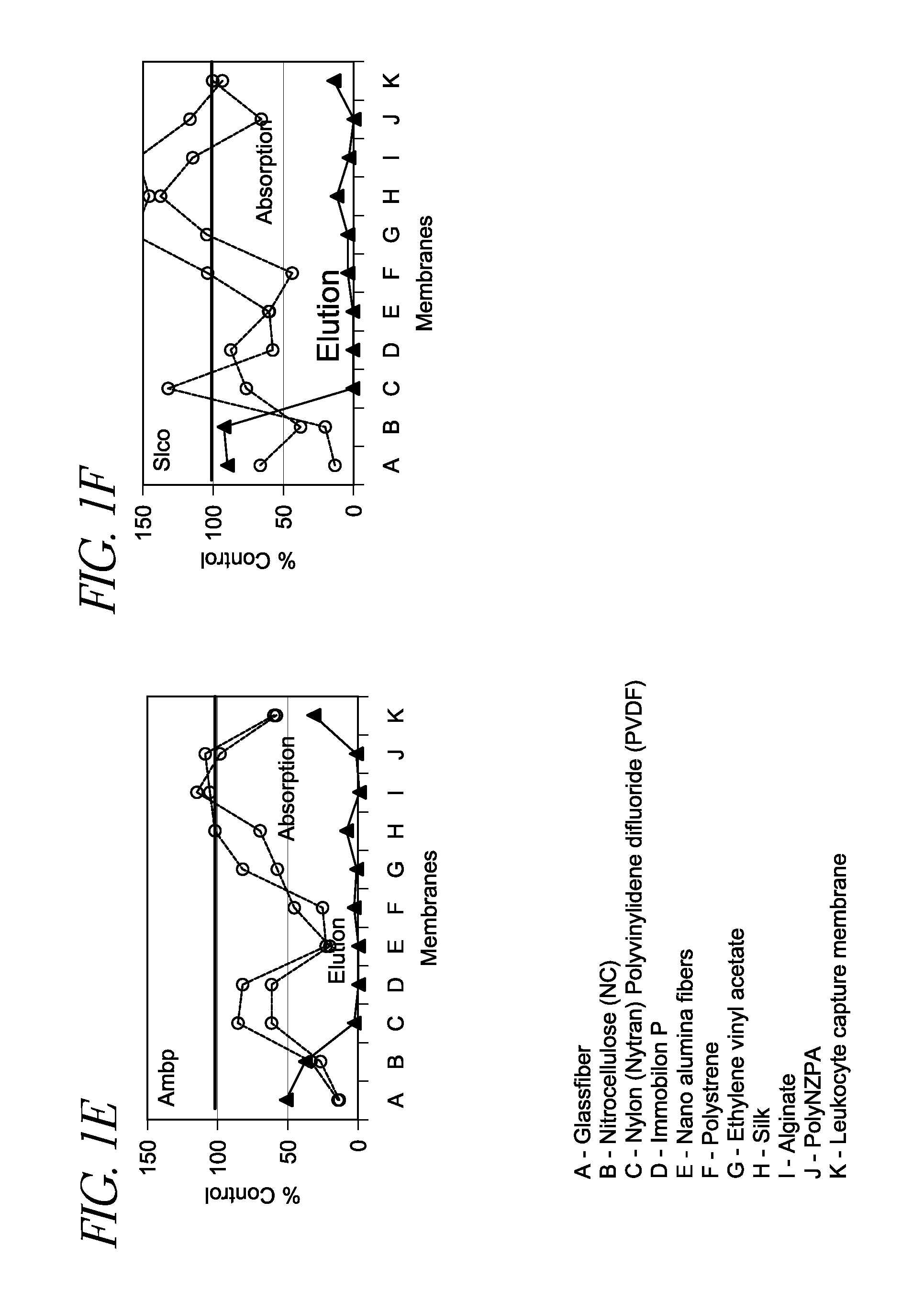
[0045]One hundred (100) μL of rat plasma was applied to a vesicle-capture device as described herein in duplicate, where various vesicle capture materials (A-K shown in FIG. 1) were inserted at the bottom of each sample receiving well. The passing through fraction was collected and mixed with 2× Lysis Buffer. The resultant lysate was subject to poly(A)+ RNA preparation and cDNA synthesis using an oligo(dT)-immobilized microplate. Further information regarding the oligo(dT)-immobilized microplate can be found in U.S. Pat. No. 7,745,180, issued on Jun. 29, 2010, and Mitsuhashi et al., Quantification of mRNA in whole blood by assessing recovery of RNA and efficiency of cDNA synthesis. Clin. Chem. 52:634-642, 2006, the disclosure of each of which is incorporated be reference herein. Various mRNAs (group-specific component (gc), apolipoprotein H (apoh), solute carrier organic anion transporter family member 1b2 (s1c1b2), plasminogen (plg),...
example 3
apture from Saliva
[0048]Ten, 1000 μL, saliva samples from a single donor were applied to a vesicle-capture device as described comprising glassfiber filters. After centrifugation at 2,000×g for 5 min, Lysis Buffer was applied and incubated at 55° C. for 30 min. Lysate was then transferred to an oligo(dT)-immobilized microplate for mRNA purification, followed by cDNA synthesis and real time PCR, according to protocol discussed above and further described in U.S. Pat. No. 7,745,180. As shown in FIG. 3, various mRNAs (ACTB, B2M, IL8, and trypsin) were detected from saliva. Consequently, the filter-based methods and devices disclosed herein are capable of capturing exosomes from saliva samples without the need for costly ultracentrifugation and with decreased risk of contamination (due to the more streamlined protocol).
example 4
tion of Captured Materials on Glassfiber Filter
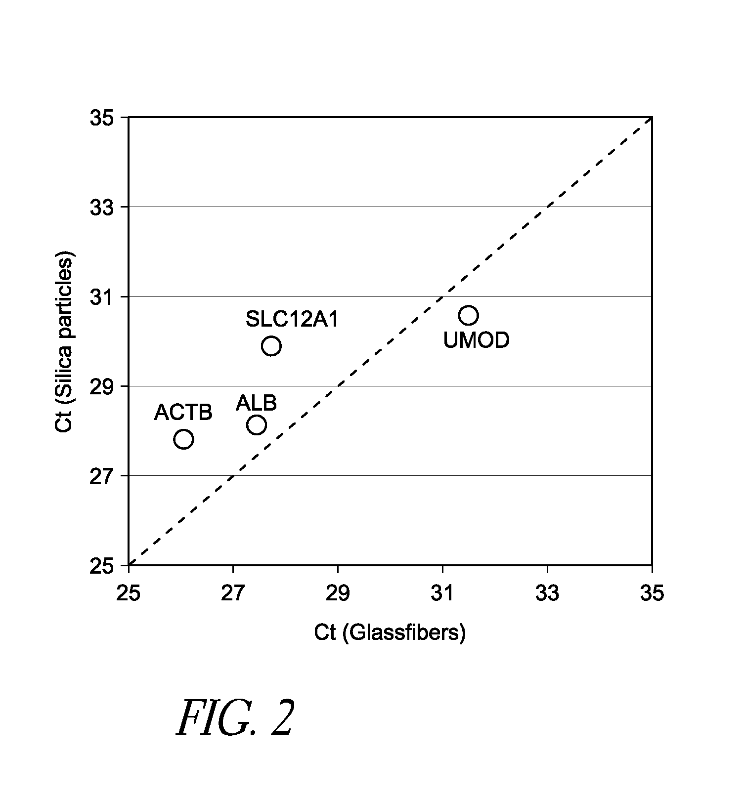
[0049]Human urine was applied to a glassfiber filter and subsequently washed with water. Vesicle-laden filters were then analyzed by scanning electron microscope (SEM). As shown in FIG. 4, small vesicle-like materials were trapped by the mesh of filters (white arrows in 4A and 4B). Vesicles also adhered to the fiber surfaces (white arrows in 4C and 4D). Occasionally, the aggregates of vesicles were also trapped by the filter (white arrows in 4E and 4F). According to the method discussed above, it was confirmed that this filter at least contained β-actin (ACTB), solute carrier family 12A1 (SLC12A1), and uromodulin (UMOD) mRNA, which are all markers that could be used as diagnostic or control markers from a urine sample. Thus, the devices and methods disclosed herein are well suited for capture of vesicles from urine samples without the need for costly ultracentrifugation and with decreased risk of contamination.
Example 5—Scanning Electro...
PUM
| Property | Measurement | Unit |
|---|---|---|
| diameter | aaaaa | aaaaa |
| size | aaaaa | aaaaa |
| diameter | aaaaa | aaaaa |
Abstract
Description
Claims
Application Information
 - R&D
- Intellectual Property
- Life Sciences
- Materials
- Tech Scout
- Unparalleled Data Quality
- Higher Quality Content
- 60% Fewer Hallucinations
Browse by: Latest US Patents, China's latest patents, Technical Efficacy Thesaurus, Application Domain, Technology Topic, Popular Technical Reports.
© 2025 PatSnap. All rights reserved.Legal|Privacy policy|Modern Slavery Act Transparency Statement|Sitemap|About US| Contact US: help@patsnap.com


