
Method for observing protein crystal

Inactive Publication Date: 2013-07-18
OSAKA UNIV
View PDF0 Cites 1 Cited by
  • Summary
  • Abstract
  • Description
  • Claims
  • Application Information

AI Technical Summary

Benefits of technology

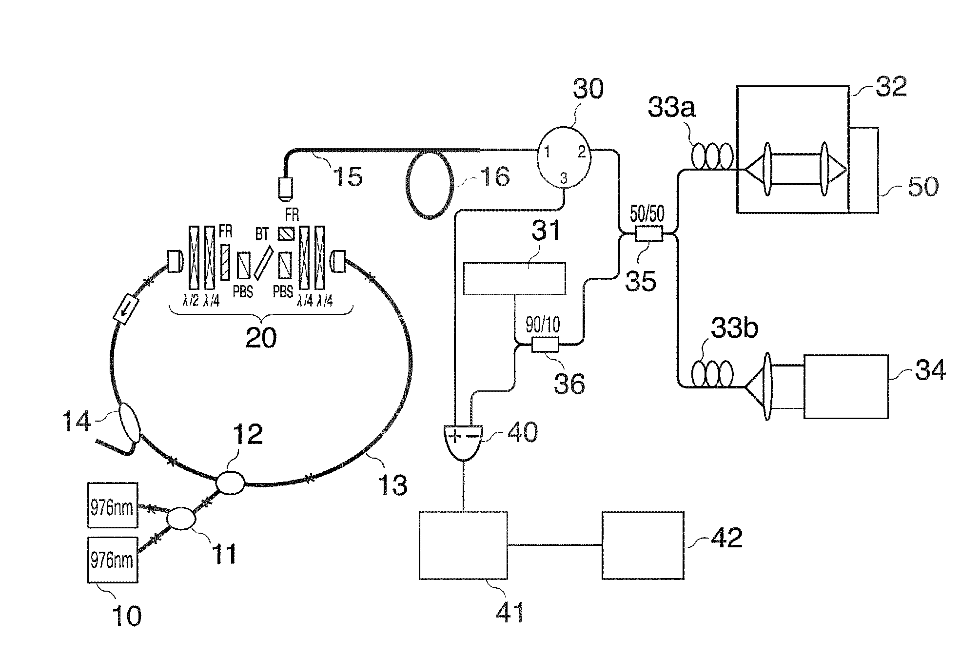
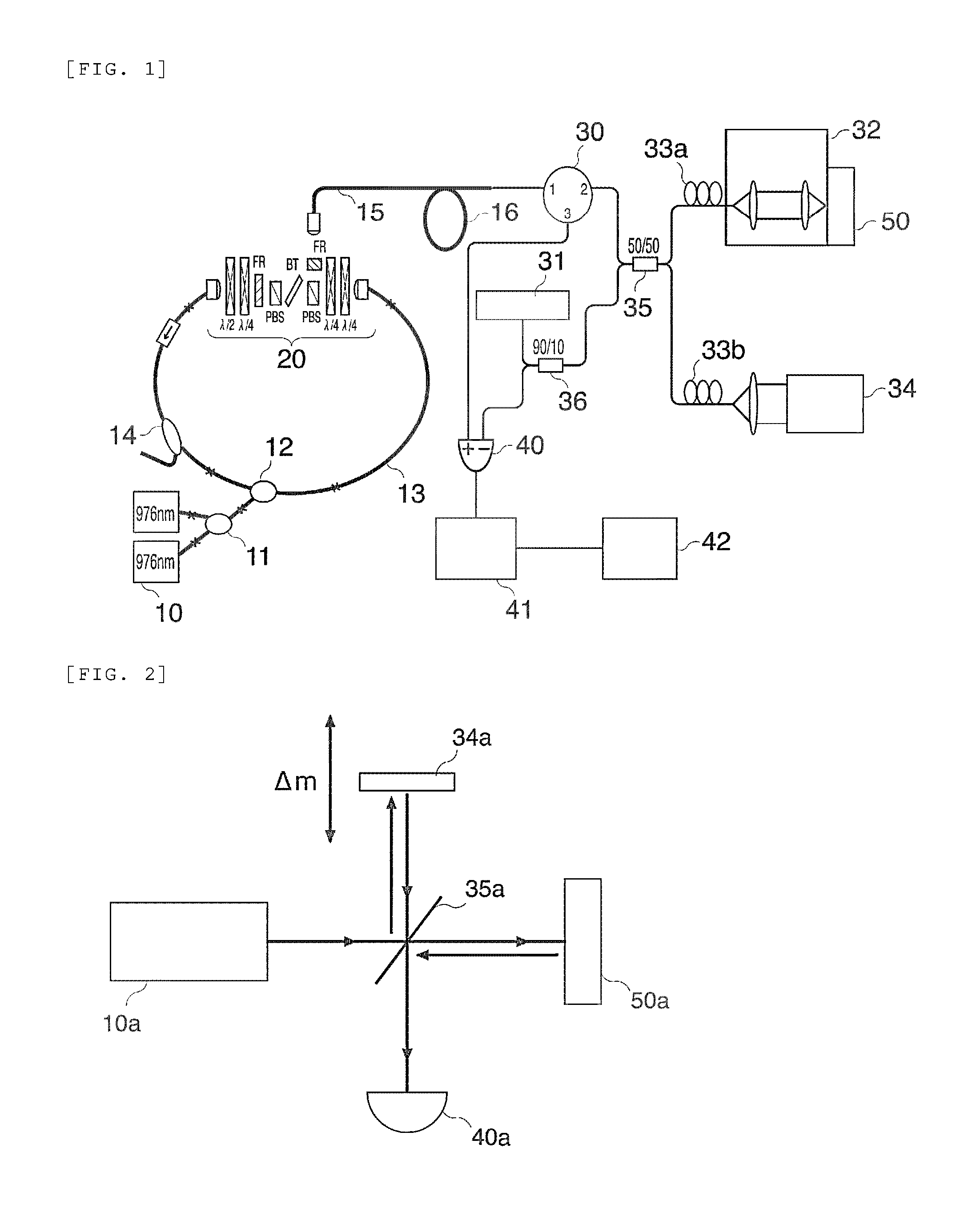
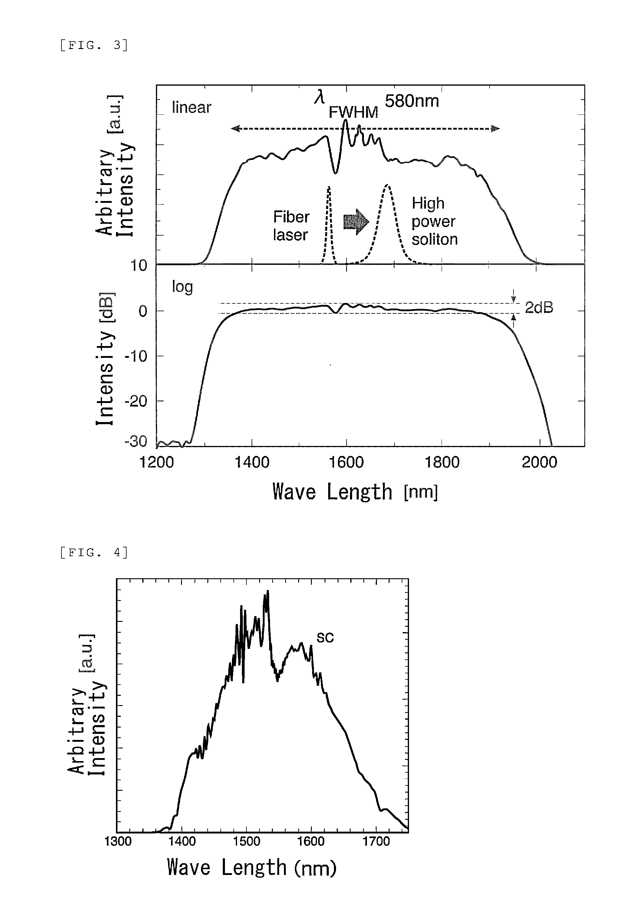
The present invention allows for the three-dimensional observation of protein crystal growth in real-time without destruction, resulting in precise control of crystal growth and the production of high-quality single crystals. The invention is achieved through the use of an OCT device that utilizes ultra-wideband scalable coherent light (SC light) to generate images of the crystal growth process. The measurement theory of OCT is applied to accurately measure the intensity and wavelength of the ultra-wideband SC light in relation to the crystal growth process. The OCT device includes an ultra-wideband SC light generating part, which is designed to generate high-quality images of crystal growth.

Problems solved by technology

Meanwhile, regarding provision of protein crystals to be subjected to this X-ray crystal analysis, it is hard to say that environments are sufficiently regulated under present circumstances.
However, there remain problems of the presence of a protein crystal not stained, necessity of several hours for staining, unusability of a crystal once stained for analysis, and the like.
However, it was difficult to observe a shape along the depth direction (three-dimensional observation).
Observation of a crystal in a highly turbid solution was more difficult.

Method used


Image

  • Method for observing protein crystal
  • Method for observing protein crystal
  • Method for observing protein crystal

Examples


example 1

[0113]In the present example, a protein crystal grown in a gel was observed using an optical microscope and an OCT device utilizing ultra-wideband SC light as a light source, and the observation results of them were compared.

1. Crystallization Condition

(1) Preparation of Protein Solution

[0114]Egg-white lysozyme (60 mg) was dissolved in 1.0 ml of 0.1 M sodium acetate to prepare a 60 mg / ml protein solution.

(2) Preparation of Reservoir Solution

[0115]A sodium acetate solution (solvent: ultrapure water) having a concentration of 0.1 M was prepared (pH: 4.5), and further, sodium chloride was dissolved in this so as to have its concentration of 5.12 M to prepare a reservoir solution.

(3) Preparation of Agar Liquid (Gel)

[0116]Agar liquid was prepared using 3 mg of agar and 50 ml of ultrapure water. Thereafter, to 400 μl of the agar liquid was added 100 μl of ultrapure water to give gel liquid.

(4) Preparation of Sodium Citrate Solution

[0117]A sodium citrate solution having a concentration of ...

example 2

[0136]In the present example, a sample containing a protein crystal and a low molecular salt was prepared, and the observation results with an optical microscope and with an OCT device were compared.

1. Crystallization Condition

(1) Preparation of Protein Solution

[0137]Egg-white lysozyme (72 mg) was dissolved in 1.0 ml of 0.1 M sodium acetate to prepare a 72 mg / ml protein solution.

(2) Preparation of Reservoir Solution

[0138]A sodium acetate solution (solvent: ultrapure water) having a concentration of 0.1M was prepared (pH: 4.5), and further, sodium chloride was dissolved in this so as to have its concentration of 5.12 M to prepare a reservoir solution.

(3) Preparation of Agar Liquid (Gel)

[0139]Agar liquid was prepared using 3 mg of agar and 50 ml of ultrapure water. The agar liquid was used as gel liquid.

(4) Preparation of Potassium Phosphate Solution

[0140]A potassium phosphate solution sodium having a concentration of 0.6 M (solvent: ultrapure water) was prepared (pH7.5).

(5) Preparati...

example 3

[0152]In the present example, the state of crystal growth in a gel was observed regarding an Egg-white lysozyme crystal as a protein crystal while changing gel concentration.

1. Crystallization Condition

(1) Preparation of Protein Solution

[0153]Egg-white lysozyme (150 mg) was dissolved in 1.0 ml of 0.1 M sodium acetate to prepare a 150 mg / ml protein solution.

(2) Preparation of Reservoir Solution

[0154]A sodium acetate solution (solvent: ultrapure water) having a concentration of 0.1 M was prepared (pH: 4.5), and further, sodium chloride was dissolved in this so as to have its concentration of 1.53 M to prepare a reservoir solution.

(3) Preparation of Agar Liquid (Gel)

[0155]Agar liquid was prepared using 3 mg of agar and 50 ml of ultrapure water. Thereafter, to 450 μl of the agar liquid was added 50 μl of ultrapure water to give gel liquid.

(4) Production of Protein Crystal

[0156]The gel liquid (2 μl), the protein solution (2 μl) and the reservoir solution (2 μl) were mixed, to obtain a cr...


PUM

PropertyMeasurementUnit
Wavelengthaaaaaaaaaa

Abstract

Provided are a method for observing protein crystal, wherein the growth process of the protein crystals is nondestructively and three-dimensionally monitored on a real-time basis and the growth of the crystals is controlled at a high accuracy to thereby enable the formation of single crystals having good qualities, which comprises observing the protein crystals, said protein crystals having been produced by a crystallization method using a gel, by an OCT measurement using light emitted from an ultrawideband light source;a method for observing protein crystals wherein the ultrawideband light source is an ultrawideband supercontinuum light source;a method for observing protein crystals wherein the center wavelength of the light emitted from the ultrawideband supercontinuum light source is a 0.8 μm band; anda method for observing protein crystals wherein the monitoring of the protein crystals is a monitoring by an in situ measurement.

Description

TECHNICAL FIELD[0001]The present invention relates to a method for observing protein crystal for producing a good quality single crystal of a protein.BACKGROUND ART[0002]Now a day decoding of human genome has been completed and the steric structures of disease-related proteins as drug targets are being distinguished one after another by a proteomic study. By analysis of the steric structures of these disease-related proteins, it becomes possible to design a compound controlling the function of a protein, thus, drug discovery processes can be shortened significantly.[0003]Therefore, competitions to analyze the steric structure of a protein more quickly and with higher precision using X-ray crystal analysis have intensified.[0004]Accordingly, automatic X-ray diffraction measurement devices and the like have been developed. Further, synchrotron radiation is utilized, and large scale calculators have been developed. Thanks to the above and other, environments of analysis apparatuses are...

Claims


Application Information

Patent Timeline
IPC IPC(8): G01B9/02C07K1/30
CPCG01N21/4795G01B9/02091C07K1/306G01N2021/1787
Inventor NISHIZAWA, NORIHIKOMATSUMURA, HIROYOSHIMORI, YUUSUKEISHIDA, SHUTAROITOH, KAZUYOSHISUGIYAMA, SHIGERUADACHI, HIROAKIINOUE, TSUYOSHITAKANO, KAZUFUMISATOSHI, MURAKAMI
Owner OSAKA UNIV
Learn More