
Device for obtaining stereoscopic images from a conventional endoscope with single lens camera

a technology of stereoscopic images and endoscopes, which is applied in the field of devices for obtaining stereoscopic images from a conventional endoscope with single lens camera, can solve the problems of prohibitive introduction into the body cavity, difficult to perform delicate operations, and large size of the endoscope having two optic channels,

Inactive Publication Date: 2012-10-04
SOOD SANDEEP +1
View PDF3 Cites 5 Cited by
  • Summary
  • Abstract
  • Description
  • Claims
  • Application Information

AI Technical Summary

Benefits of technology

The present invention allows for stereoscopic viewing with existing endoscopes by eliminating the requirement for a special lens system or adding any new equipment between the endoscope and camera. Instead, a unique method uses a two-dimensional lens scope and requires no modification whatsoever to its structure. Only one image path is needed to create a realistic and immersive experience for medical professionals during procedures.

Problems solved by technology

The technical problem addressed in this patent is how to convert a regular endoscope into a stereoscopic device without being too heavy or bulky, while still achieving proper alignment and color balance between the left and right eyes. Previous solutions involved expensive and complicated modifications to the endoscope or added extra components like duel lenses or specialized adapters.

Method used


Image

  • Device for obtaining stereoscopic images from a conventional endoscope with single lens camera
  • Device for obtaining stereoscopic images from a conventional endoscope with single lens camera
  • Device for obtaining stereoscopic images from a conventional endoscope with single lens camera

Examples


Embodiment Construction

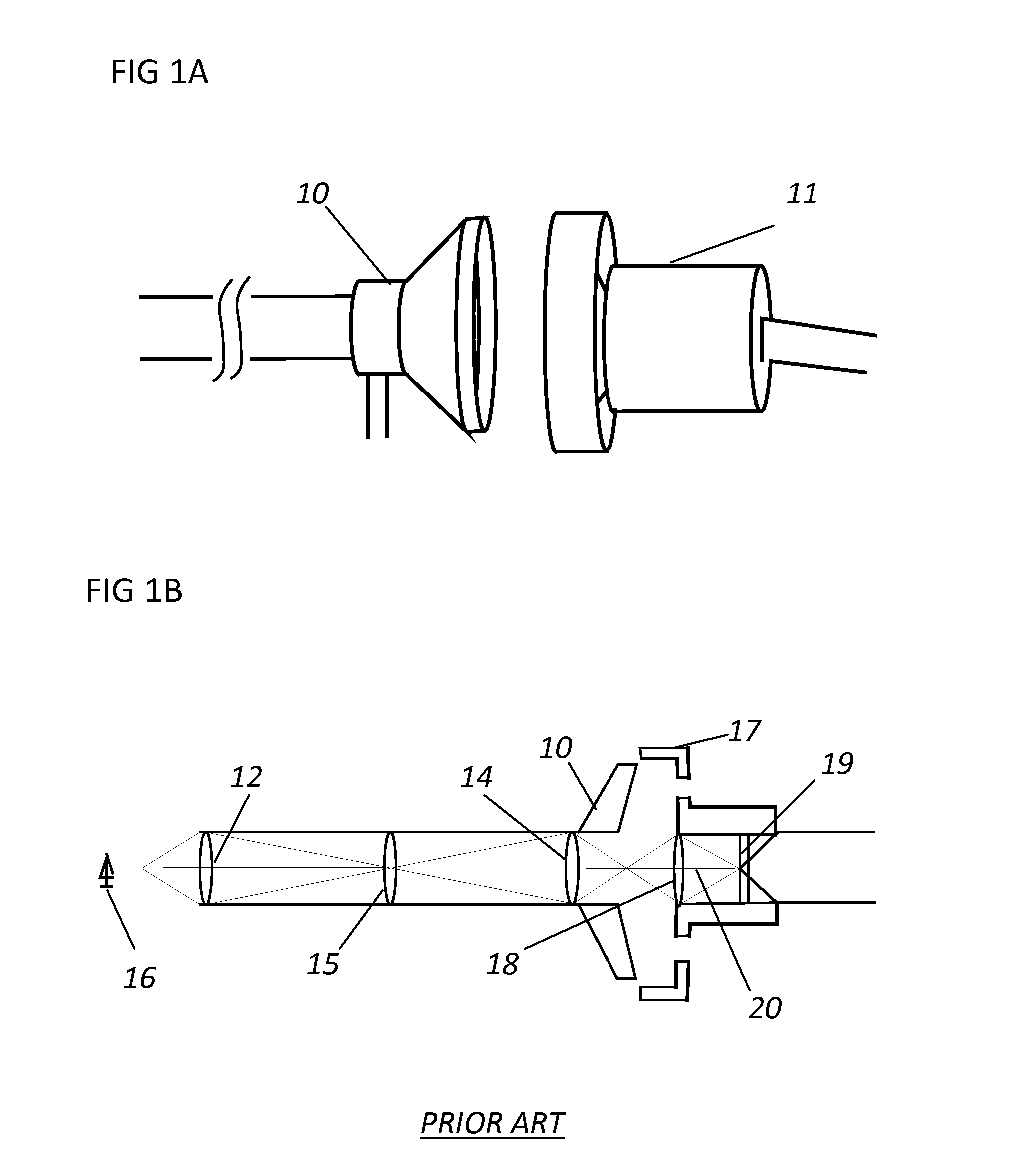
[0032]FIG. 1A illustrates a known type of endoscope 10 for two dimensional viewing using a camera 11. In schematic FIG. 1B as shown, the endoscope 10 has entry lenses 12 and exit lenses 14 with other lens 15 there between. Rays from an object 16 pass through the entry pupil and entry lens 12. A coupler 17 couples the endoscope to the camera 11. The rays are focused by a focusing lens 18 on the light sensitive element 19 of the camera 11. The principal ray 20 is shown at the central axis of the endoscope 10.

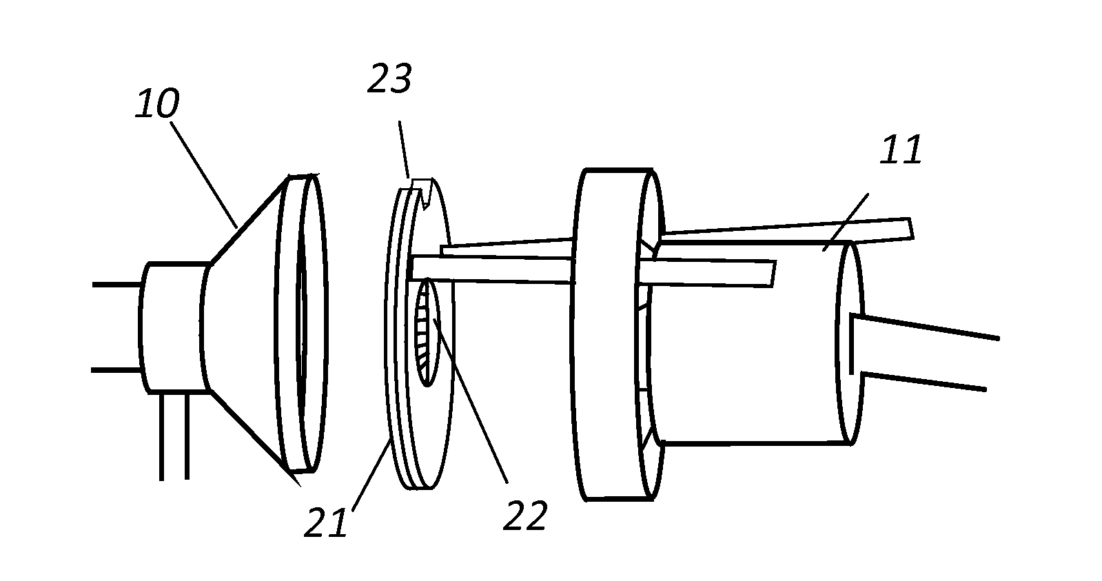
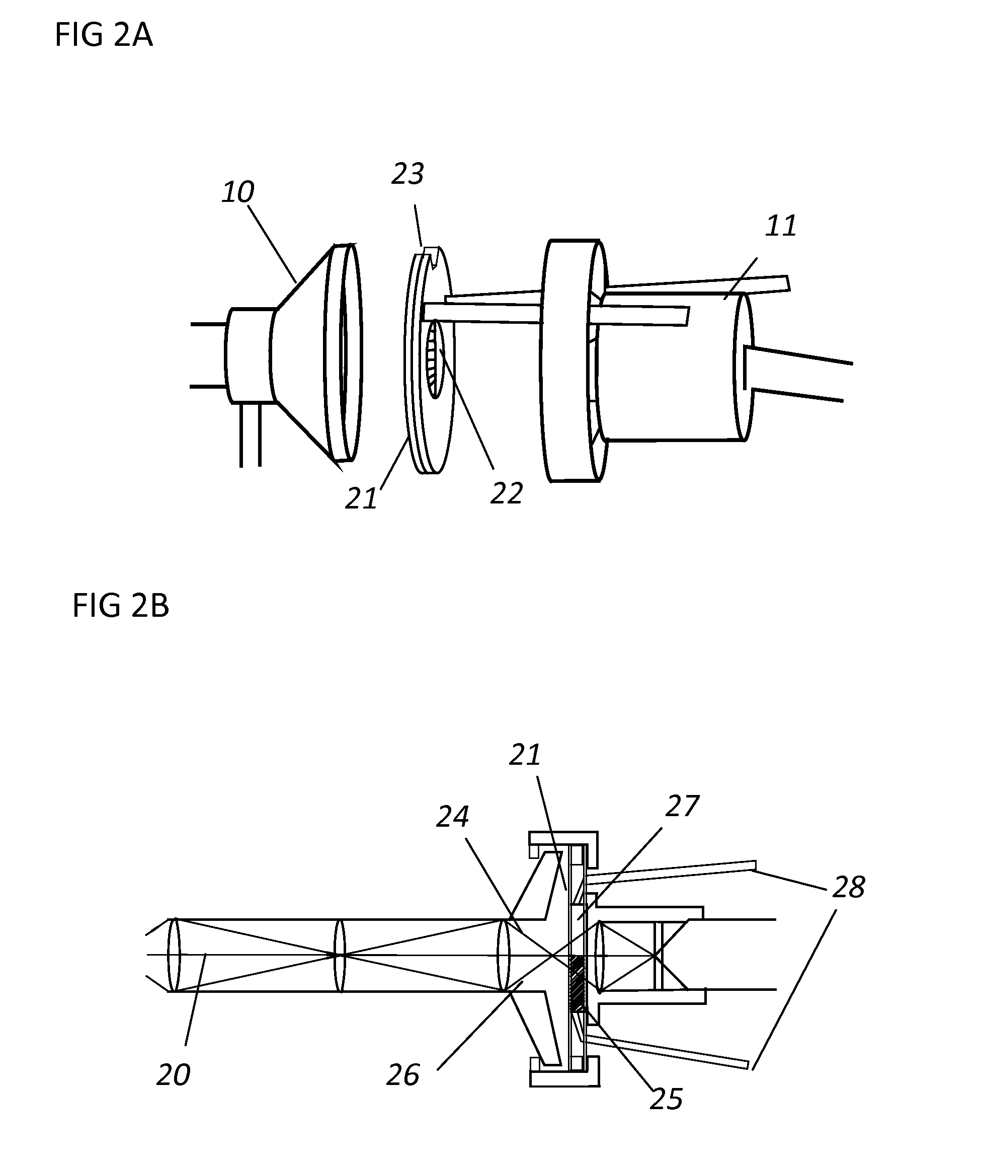
[0033]FIG. 2A shows the endoscope 10 of FIG. 1 modified for stereoscopic viewing system by placing the device 21 with an aperture 22, disclosed herewith, between the endoscope 10 and the camera 11 with notch 23 on the device positioned at the top. As in FIG. 2B, the left perspective ray 24 passes through the left filter 25 and the right perspective ray 26 passes through the right filter 27 of the device 21. Thus, the camera sees a different perspective view of the object in the si...


PUM


Abstract

The present invention discloses a device for obtaining stereoscopic images with conventional single optic channel endoscope using a single lens camera. The device comprises of an aperture each half of which is covered with complimentary additive and subtractive color filters and is installed between the endoscope and camera. The filters can be transversely adjusted to center on the principal ray so as to obtain optimal stereopsis. Images are viewed on a standard monitor using eyewear comprising the same complimentary additive and subtractive color filters on the left and the right side, or on specialized video monitors using active or passive polarized glasses.

Description


Claims


Application Information

Patent Timeline
Owner SOOD SANDEEP
Learn More