
Ultrasonic diagnostic apparatus and region-of-interest

a diagnostic apparatus and ultrasonic technology, applied in the field of ultrasonic diagnostic apparatus and region-of-interest setting method, can solve the problem of inability to perform effective image diagnosis, and achieve the effect of improving the accuracy of roi setting in imt measuremen

Inactive Publication Date: 2012-05-24
HITACHI MEDICAL CORP
View PDF3 Cites 12 Cited by
  • Summary
  • Abstract
  • Description
  • Claims
  • Application Information

AI Technical Summary

Benefits of technology

[0012]The present invention is effective in providing the ultrasonic diagnostic apparatus and the region setting method capable of improving accuracy of ROI setting in IMT measurement.BRIEF DESCRIPTION OF THE DIAGRAMS
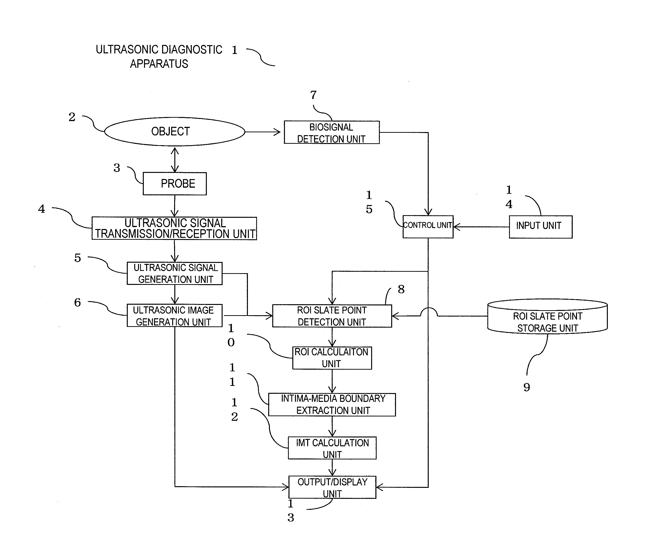
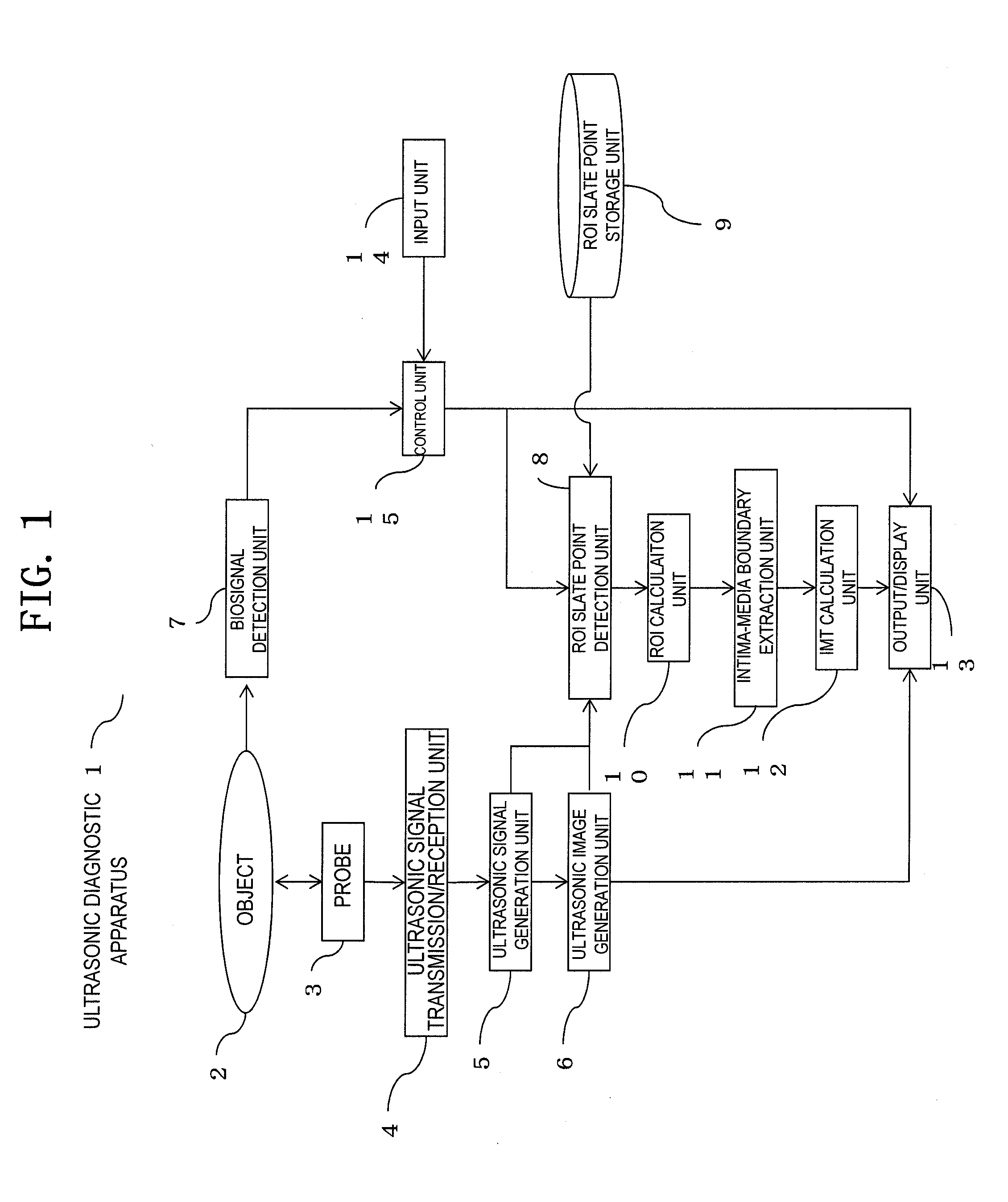
[0013]FIG. 1 is a block diagram showing a configuration example of the ultrasonic diagnostic apparatus in a first embodiment of the present invention.
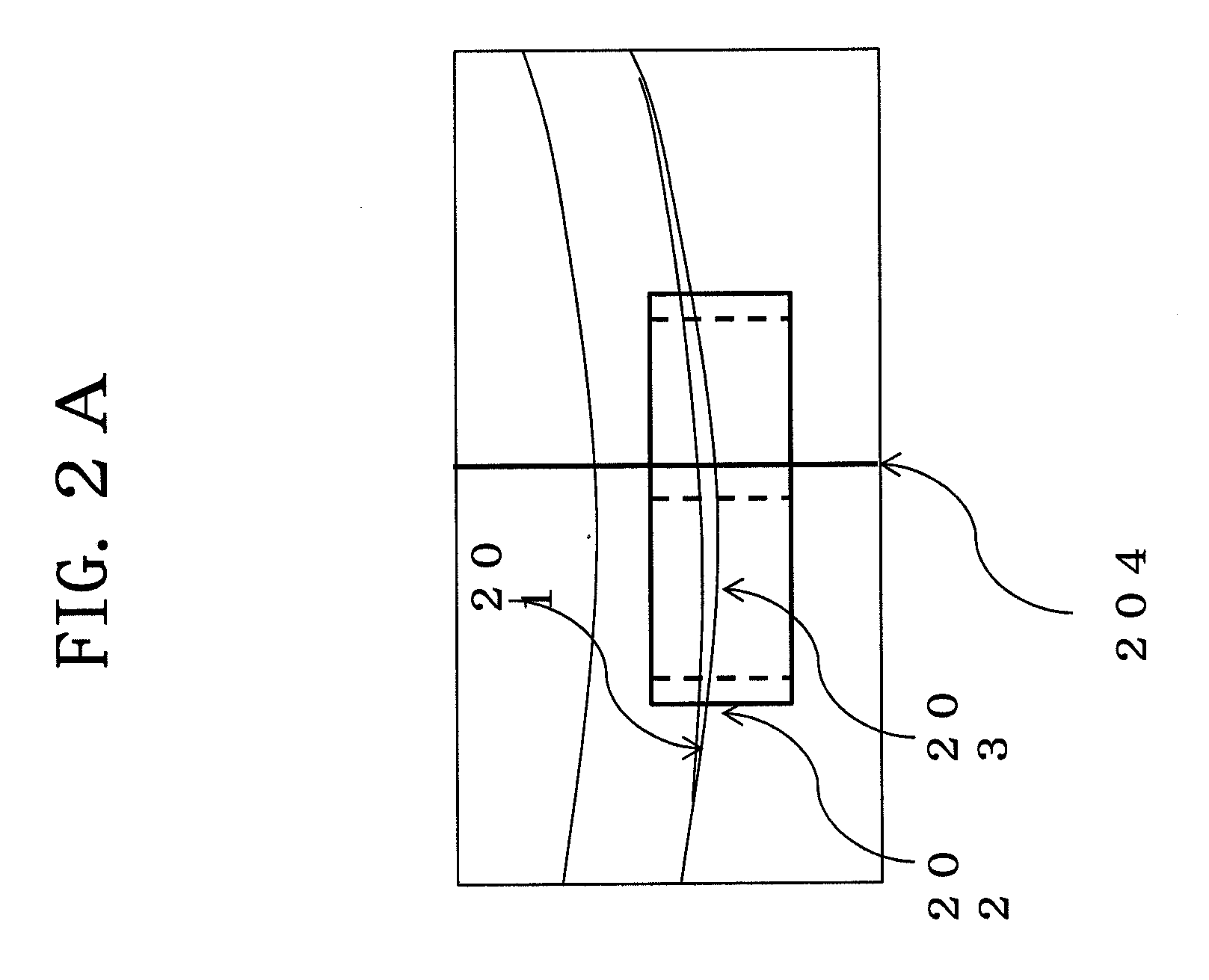
[0014]FIG. 2A is a view for explaining the characteristic of brightness change in a carotid wall.
[0015]FIG. 2B is a view for explaining brightness change on a line 204 in FIG. 2A.
[0016]FIG. 3 is a view for explaining the principle of ROI setting in the first embodiment of the present invention.
[0017]FIG. 4 is a flowchart for explaining an operation example of the ultrasonic diagnostic apparatus in the first embodiment of the present invention.

Problems solved by technology

However, if an examiner executes manual ROI setting operation related to IMT measurement for all of patients, effective image diagnosis cannot be performed.

Method used


Image

  • Ultrasonic diagnostic apparatus and region-of-interest
  • Ultrasonic diagnostic apparatus and region-of-interest
  • Ultrasonic diagnostic apparatus and region-of-interest

Examples


embodiment 1

[0024]FIG. 1 is a block diagram showing the outline of the ultrasonic diagnostic apparatus in the first embodiment of the present invention.

[0025]In the first embodiment, an ultrasonic probe 3, an ultrasonic transmission / reception unit 4, an ultrasonic signal generation unit 5 and an ultrasonic image generation unit 6 take on the function to “execute imaging of an ultrasonic image by transmitting / receiving ultrasonic waves to / from a region including a carotid artery of an object”.

[0026]Also, an ROI slate point detecting unit 8, an ROI slate point storing unit 9 and ROI calculating unit 10 take on the function to “scan the ultrasonic image and set an ROI region including the intima-media complex on the ultrasonic image based on the degree of concentration of contour slate points of the carotid artery by a region-of-interest setting unit”.

[0027]Also, an intima-media complex contour extracting unit 11 and an IMT calculating unit 12 take on the function to “measure intima-media thicknes...

embodiment 2

[0110]The second embodiment exemplifies the case that there are two or more ROIs. Since the configuration and operation of the ultrasonic diagnostic apparatus 1 is the same as the first embodiment, the description thereof will be omitted and only different parts will be described.

[0111]The calculation step of the position and the size of an ROI will be described referring to FIG. 6.

[0112]FIG. 6 is a view explaining the principle of ROI setting in the second embodiment of the present invention.

[0113]First, slate data 603 is plotted on the ultrasonic image in which the carotid artery to be displayed on the image display unit 502 in a screen is depicted. Then plural sets of the slate point data 603 are acquired by the same procedure, and plotted in the same manner on the ultrasonic image. The coordinate points of the plotted slate point data 603 on the ultrasonic image are stored to be read out in the subsequent process.

[0114]Next, the control unit 15 scans the pixel points of the ultr...

embodiment 3

[0126]The third embodiment explains an example that the ROI setting executed on the blood vessel wall of a carotid artery which is closer to an ultrasonic probe (one side) is reflected on the ROI setting of the blood vessel which is farther from the probe (the other side).

[0127]Since the configuration and operation of the ultrasonic diagnostic apparatus 1 is the same as the first embodiment, the description thereof will be omitted and only the parts different from the first embodiment will be described.

[0128]The calculation step of the position and the size of an ROI in the present embodiment will be described using FIG. 7.

[0129]FIG. 7 is a view for explaining the principle of ROI setting in the third embodiment of the present invention.

[0130]First, data processing is executed on one side of the carotid artery which is an outer wall part in the lower side of the diagram, as in FIG. 3.

[0131]Next, the following data processing is executed on the other side of the carotid artery which ...


PUM


Abstract

Disclose is an ultrasonic diagnostic apparatus which transmits / receives an ultrasonic wave to / from a region including a carotid artery of an object, images an ultrasonic image, and comprises a thickness measurement unit configured to measure the thickness of an intima-media complex from the ultrasonic image, the ultrasonic diagnostic apparatus being provided with a region-of-interest setting unit configured to scan the ultrasonic image and set a region of interest including the intima-media complex on the ultrasonic image on the basis of the degree of concentration of contour slate points of the carotid artery, wherein the thickness measurement unit measures the thickness of the intima-media complex on the basis of boundaries within the set region of interest.

Description

FIELD OF THE INVENTION[0001]The present invention relates to an ultrasonic diagnostic apparatus and region-of-interest setting method therefore capable of setting a region of interest (ROI) which is a target for measuring intima-media thickness (IMT) in an ultrasonic image of a carotid artery of an imaged object so as to measure an IMT value.DESCRIPTION OF RELATED ART[0002]IMT measurement is known as effective for finding arterial sclerosis in an object to be examined. IMT measurement requires the procedure that a medical service worker (an examiner) such as a doctor or a clinical laboratory technologist obtains an ultrasonic image by applying an ultrasonic probe onto a carotid artery part of the object and sets an ROI on the obtained ultrasonic image.[0003]The examiner may set an ROI manually. However, if an examiner executes manual ROI setting operation related to IMT measurement for all of patients, effective image diagnosis cannot be performed.[0004]Given this factor, Patent Doc...

Claims


Application Information

Patent Timeline
Patent Type & Authority Applications(United States)
IPC IPC(8): A61B8/00G06K9/00
CPCA61B8/02A61B8/06A61B8/0891G06T2207/30101A61B8/5223G06T7/602G06T2207/10132A61B8/469G06T7/62G16H50/30
Inventor CHONO, TOMOAKI
Owner HITACHI MEDICAL CORP
Learn More