Near infra red fluorescence imaging for visualization of blood vessels during endoscopic harvest

a technology of endoscopic harvest and which is applied in the field of near infra red fluorescence imaging for visualization of blood vessels during endoscopic harvest, can solve the problems of trauma to the vessel, difficulty in recognizing the side branches of the vessel by white light imaging, etc., and achieve the effect of reducing/preventing the potential of vessel trauma

Active Publication Date: 2010-11-11
STRYKER EUROPEAN OPERATIONS LIMITED
View PDF5 Cites 33 Cited by
  • Summary
  • Abstract
  • Description
  • Claims
  • Application Information

AI Technical Summary

Benefits of technology

[0010]In another embodiment, the vessel tortuousity and the presence of side-branches is evaluated to reduce / prevent the potential of vessel trauma and subsequent related adverse outcomes.

Problems solved by technology

While yet to be definitively ascertained, this adverse outcome is thought to be associated with vessel trauma incurred during the endoscopic harvest procedure.
Following the course of the vessel during dissection and recognizing of side branches of this vessel by white light imaging is often problematic.
Difficulties in recognizing the course of the vessel and / or identifying side branches may lead to trauma to the vessel, with the potential for sub optimal patient outcome.

Method used


Image

  • Near infra red fluorescence imaging for visualization of blood vessels during endoscopic harvest
  • Near infra red fluorescence imaging for visualization of blood vessels during endoscopic harvest
  • Near infra red fluorescence imaging for visualization of blood vessels during endoscopic harvest

Examples


Embodiment Construction

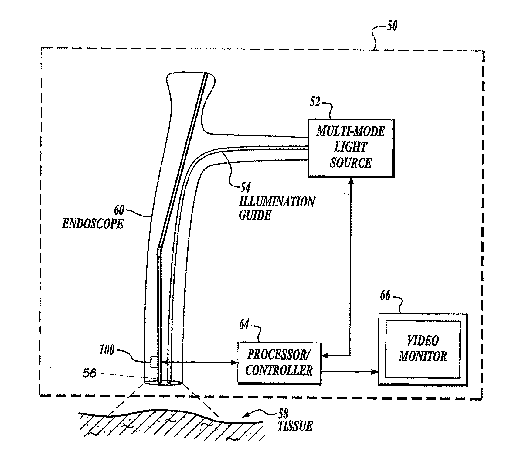
[0017]The invention is directed to preoperative / intra-operative determination of the location of vessels to be harvested endoscopically and to enhance visualization of them of them throughout the procedure.

[0018]The system includes a multi-mode light source 52 that generates light for obtaining color and fluorescence images. The use of the light source for obtaining different kinds of images will be described in further detail below. Light from the light source 52 is supplied to an illumination guide 54 of an endoscope 60, which then illuminates a tissue sample 58 that is to be imaged. The system also includes a camera 100 located at the insertion end of the endoscope 60. The light from the tissue is directly captured by the camera 100. The endoscope 60 is similar to conventional video endoscopes, but with the added capability to provide both fluorescence / reflectance and / or fluorescence / fluorescence imaging in additional to conventional color imaging. For vessel insufflation, it als...


PUM


Abstract

Endoscopic identification and dissection of a blood vessel in tissue during endoscopic harvest can be enhanced by injecting a bolus of ICG into the bloodstream, illuminating the target vessels with excitation light capable of exciting fluorescence in the ICG, and locating the vessel to be harvested by observing the ICG fluorescence. Moreover, collapsed blood vessel may be harvested by inserting an endoscope into the collapsed blood vessel, injecting into the collapsed blood vessel a bolus containing ICG to re-inflate the blood vessel, illuminating the target vessels with excitation light capable of exciting fluorescence in the ICG; and locating the re-inflated blood vessel by observing the ICG fluorescence.

Description

CROSS-REFERENCE TO RELATED APPLICATIONS[0001]This application claims priority to U.S. Provisional Application Ser. No. 61 / 176,642, filed May 8, 2009, the entire content is incorporated herein by reference.BACKGROUND OF THE INVENTION[0002]Endoscopic Vein Harvest (EVH) has become the standard method for dissection and harvest of conduit used in coronary artery bypass surgery (CABG). Recent data show that, while overwhelmingly preferred by patients, EVH has been associated with a decrease in long-term graft patency rates. While yet to be definitively ascertained, this adverse outcome is thought to be associated with vessel trauma incurred during the endoscopic harvest procedure.[0003]Currently, identification of blood vessels to be harvested endoscopically relies upon locating the vessel by white-light visual guidance. Following the course of the vessel during dissection and recognizing of side branches of this vessel by white light imaging is often problematic. Difficulties in recogni...

Claims


Application Information

Patent Timeline
Patent Type & Authority Applications(United States)
IPC IPC(8): A61B6/00
CPCA61B1/0005A61B1/043A61B5/489A61B5/0071A61B5/0086A61B1/05
Inventor CARROLL, DOUGLAS R.FLOWER, ROBERT W.DOCHERTY, JOHN C.
Owner STRYKER EUROPEAN OPERATIONS LIMITED
Why Eureka
Try Eureka