Diagnosis and treatment of neuroectodermal tumors
a neuroectodermal tumor and tumor technology, applied in the field of cell physiology, neurology, developmental biology, oncology, can solve the problems of nestin, keratin, keratin, and glioma cell antigens that have not yet been shown to be useful for diagnostic purposes, and the gene has yet to be demonstrated in glioma cells
- Summary
- Abstract
- Description
- Claims
- Application Information
AI Technical Summary
Problems solved by technology
Method used
Image
Examples
example 1
Summary of Chlorotoxin Results from Glioma Experiments
[0072]Recent studies demonstrated a common antigen is expressed by the vast majority of glioma cells. This antigen is targeted by chlorotoxin (Ctx or TM-601), a 36 amino acid peptide originally isolated from Leiurus quinquestriatus scorpion venom. Chlorotoxin selectively binds to the membrane of glioma cells allowing selective targeting of these cells within the CNS (18). The antigen targeted by this peptide appears to be a chloride ion channel although the antigen has not yet been unequivocally identified at the molecular level. Thus far, the data indicates that chlorotoxin binds to a membrane protein of 72 kDa molecular weight that is preferentially expressed in the cytoplasmic membrane of glioma cell. Binding of the peptide enhances glioma cell proliferation (19) and inhibits the ability of glioma cells to migrate in Transwell assays, an in vitro assay to evaluate tumor invasiveness (20). Chlorotoxin appears to exert these eff...
example 2
Immunohistochemical Staining of Gliomas with Chlorotoxin
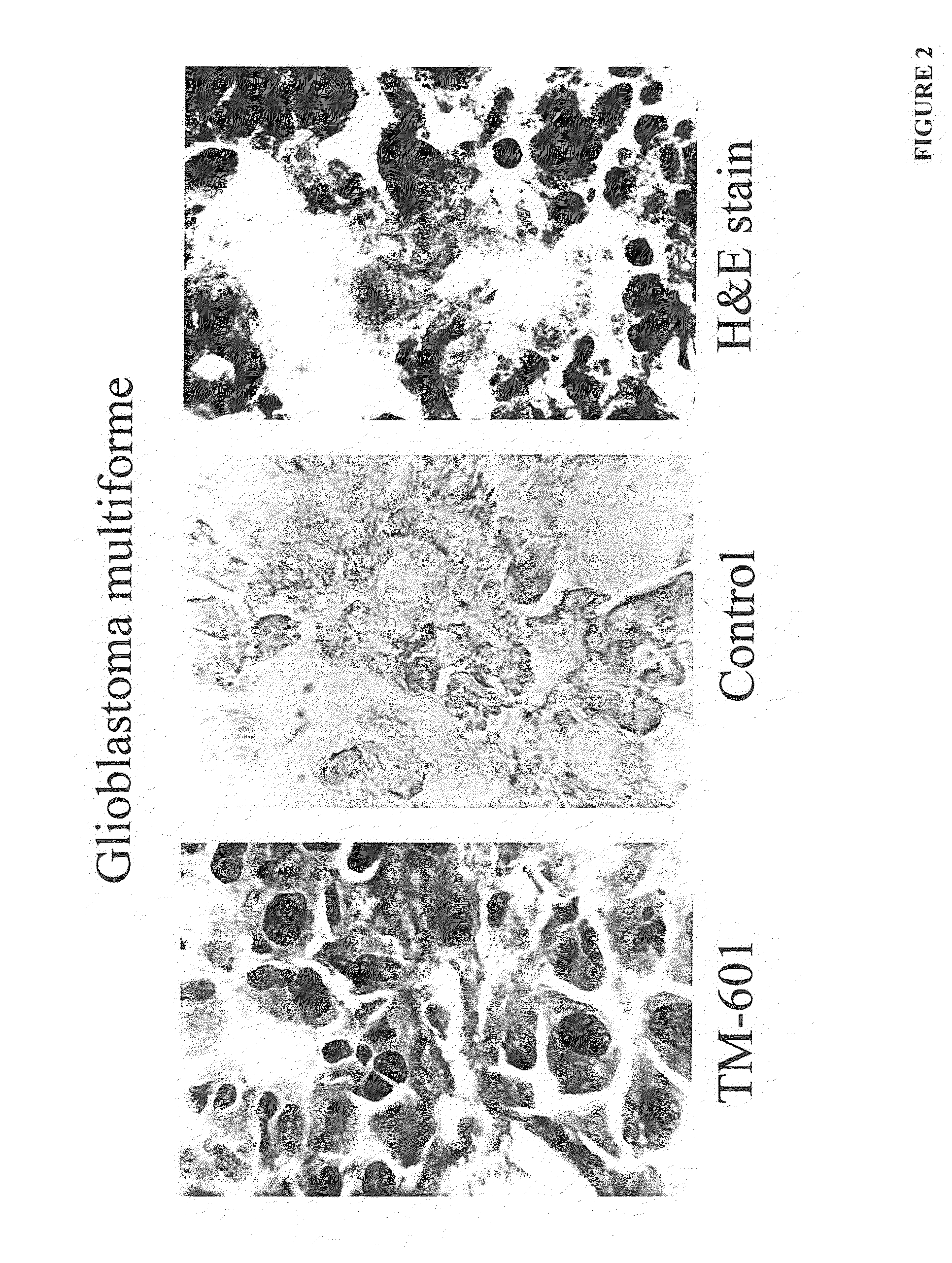
[0073]Over 250 frozen or, paraffin sections of human biopsy tissues were histochemically stained with a chemically synthesized form of chlorotoxin containing a detectable biotin group chemically attached to the N terminus (TM-601). Binding of the TM-601molecule was observed on selective cells associated with the essentially all glioma tumors with up to 95% positive cells per tumor. Based on these studies, it has been proposed to utilize chlorotoxin a s a glioma specific marker and as a potential therapeutic tool for targeting glioma tumors. For such purposes, chlorotoxin linkage of radioactive molecules or cytotoxic moieties such as saporin could be employed.
example 3
Recombinant DNA Manipulation of Chlorotoxin
[0074]Using techniques well known in the art, one may prepare recombinant proteins specifically engineered to mimic the binding and action of the native toxin. The biological activity of the synthetic chlorotoxin is as effective for chloride ion channel blockade as the native venom toxin. Recombinant techniques are used to synthesize chlorotoxin in E coli using a modified PGEX vector system and the toxin may be linked to various fusion proteins using common restriction sites. After synthesis of recombinant chlorotoxin, it may be linked to various cytotoxic fusion proteins including glutathione-S-transferase (GST), gelonin, ricin, diptheria toxin, complement proteins and radioligands and other such proteins as are well known in the immunotoxin art.
PUM
| Property | Measurement | Unit |
|---|---|---|
| molecular weight | aaaaa | aaaaa |
| fluorescent microscopy | aaaaa | aaaaa |
| fluorescent activated cell sorting | aaaaa | aaaaa |
Abstract
Description
Claims
Application Information
 - R&D
- Intellectual Property
- Life Sciences
- Materials
- Tech Scout
- Unparalleled Data Quality
- Higher Quality Content
- 60% Fewer Hallucinations
Browse by: Latest US Patents, China's latest patents, Technical Efficacy Thesaurus, Application Domain, Technology Topic, Popular Technical Reports.
© 2025 PatSnap. All rights reserved.Legal|Privacy policy|Modern Slavery Act Transparency Statement|Sitemap|About US| Contact US: help@patsnap.com


