Nucleic acids encoding recombinant 56 and 82 kDa antigents from gametocytes of Eimeria maxima and their uses
a technology of eimeria maxima and recombinant kda, which is applied in the direction of immunological disorders, drug compositions, peptides, etc., can solve the problems of coccidiosis, inability to sanitary control, and high cost of certain drugs
- Summary
- Abstract
- Description
- Claims
- Application Information
AI Technical Summary
Problems solved by technology
Method used
Image
Examples
example 1
Purification of Eimeria maxima Gametocytes on a Large Scale
[0241]In order to produce very large quantities of gametocytes, 4,000 heavy breed chickens were infected with 10,000 sporulated E. maxima oocysts, and were then sacrificed on day six (about 134 hours) post infection. The chicken intestines were removed, washed with PBS and cut open longitudinally. They were then cut into 1 cm long pieces and placed in a SAC buffered solution (170 mM NaCl, 10 mM Tris pH 7, 10 mM glucose, 5 mM CaCl2, 1% powdered milk) containing 0.5 mg / ml hyaluronidase (Type III from Sigma, 700 units / mg). The intestinal pieces were incubated at 37° C. for 20 minutes after which they were placed on top of a gauze filter. The pieces were rinsed with large quantities of SAC buffer and the resulting filtrate was collected. This was then filtered through a 17 micron polymon filter (Swiss Silk Bolting Cloth Mfg. Co. Ltd., Zurich, Switzerland) and the resulting filtrate was then filtered through a 10 micron filter. T...
example 2
Purification of the 56, 82 and 230 kDa Gametocyte Antigens
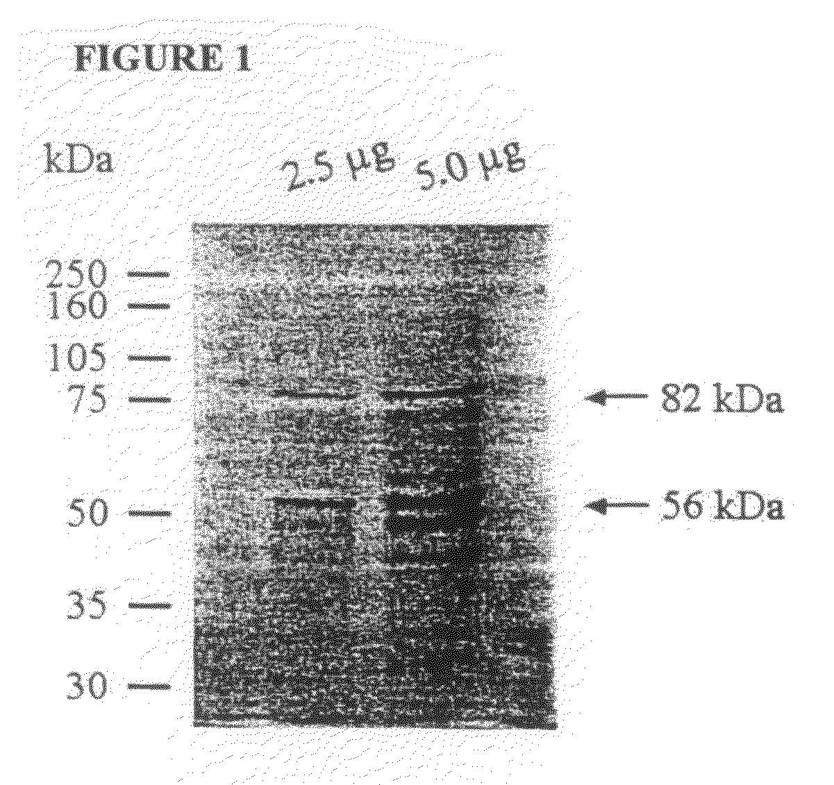
[0242]The frozen gametocytes were thawed at room temperature and the proteins were extracted as described previously (Wallach 1995). The 56 and 82 kDa gametocyte antigens were isolated from the protein extract by running it over a Sepharose 4B column containing the monoclonal antibody 1E11-11 raised against the 56 kDa antigen. A complex of the gametocyte antigens were allowed to bind to the monoclonal antibody attached to the resin, the non-specific material was washed off using buffer, and the affinity purified gametocyte antigens (APGA) were eluted from the column. The purified APGA was lyophilized. A small sample of APGA was analyzed by SDS-PAGE where the 56 and 82 kDa native antigens were clearly visualized (FIG. 1).
example 3
Two Dimensional Gel Electrophoresis of APGA and Isolation of the Major 56 and 82 kDa Antigens
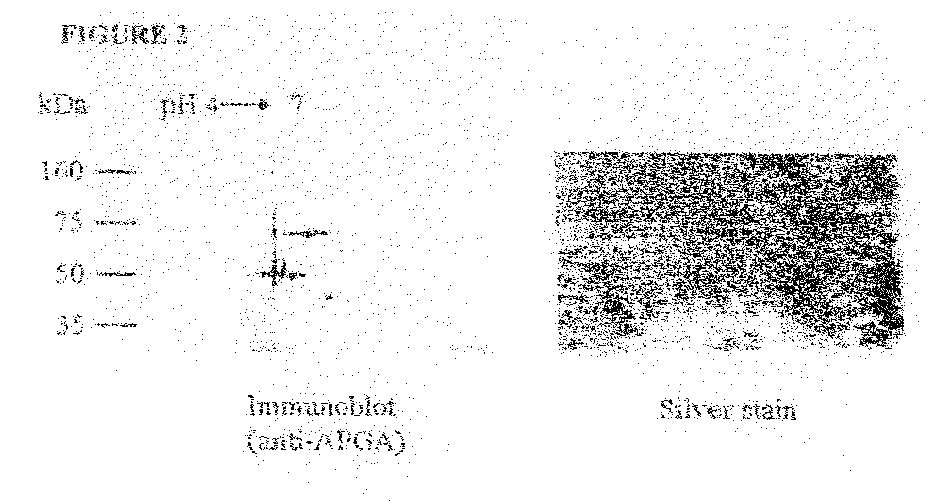
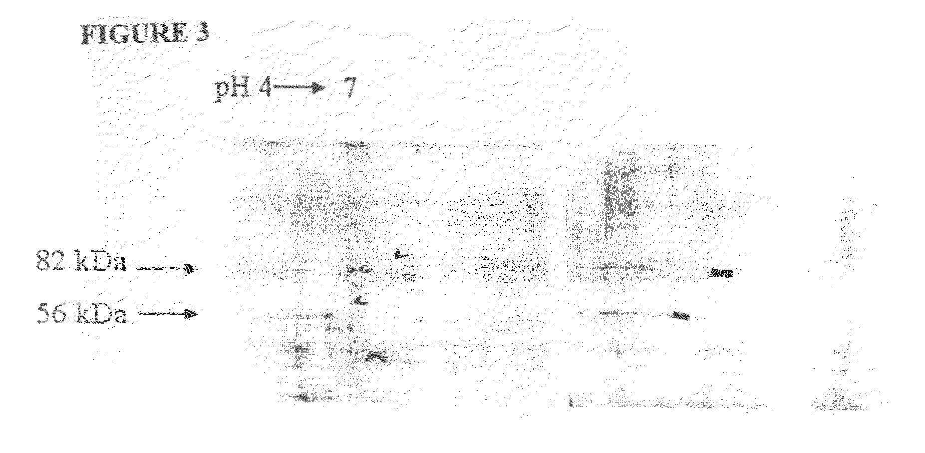
[0243]The 56 and 82 kDa gametocyte antigens were isolated from APGA. Lyophilized APGA was prepared as described in Example 2, and was solubilized in water. The proteins were then separated by two-dimensional SDS-PAGE (FIG. 2), and identified by immunoblotting using a polyclonal chicken anti-APGA antibody, which recognizes both the 56 and 82 kDa proteins. Once identified and their location established on two-dimensional SDS-PAGE gels, the proteins were then transferred to a PVDF membrane filter, and stained with Coomassie Blue (FIG. 3). Immunoblotting was carried out at the same time, and the two blots were compared to clearly identify the 56 and 82 kDa proteins. The spots corresponding to the 56 and 82 kDa gametocyte antigens were cut out of the membranes and the amino-terminus of each antigen was sequenced.
PUM
| Property | Measurement | Unit |
|---|---|---|
| molecular weights | aaaaa | aaaaa |
| molecular weights | aaaaa | aaaaa |
| molecular weights | aaaaa | aaaaa |
Abstract
Description
Claims
Application Information
 - R&D Engineer
- R&D Manager
- IP Professional
- Industry Leading Data Capabilities
- Powerful AI technology
- Patent DNA Extraction
Browse by: Latest US Patents, China's latest patents, Technical Efficacy Thesaurus, Application Domain, Technology Topic, Popular Technical Reports.
© 2024 PatSnap. All rights reserved.Legal|Privacy policy|Modern Slavery Act Transparency Statement|Sitemap|About US| Contact US: help@patsnap.com


