
Sonohysterography and biopsy catheter

a uterine abnormality and biopsy catheter technology, applied in the field of sonohysterography and biopsy catheter, can solve the problems of increasing the amount of time necessary to complete the procedure, increasing the pain of patients, increasing the recovery time of patients, etc., and achieving the effect of effective and safe procedures

Inactive Publication Date: 2008-02-21
VANCE PROD INC D B A COOK UROLOGICAL INC
View PDF4 Cites 47 Cited by
  • Summary
  • Abstract
  • Description
  • Claims
  • Application Information

AI Technical Summary

Benefits of technology

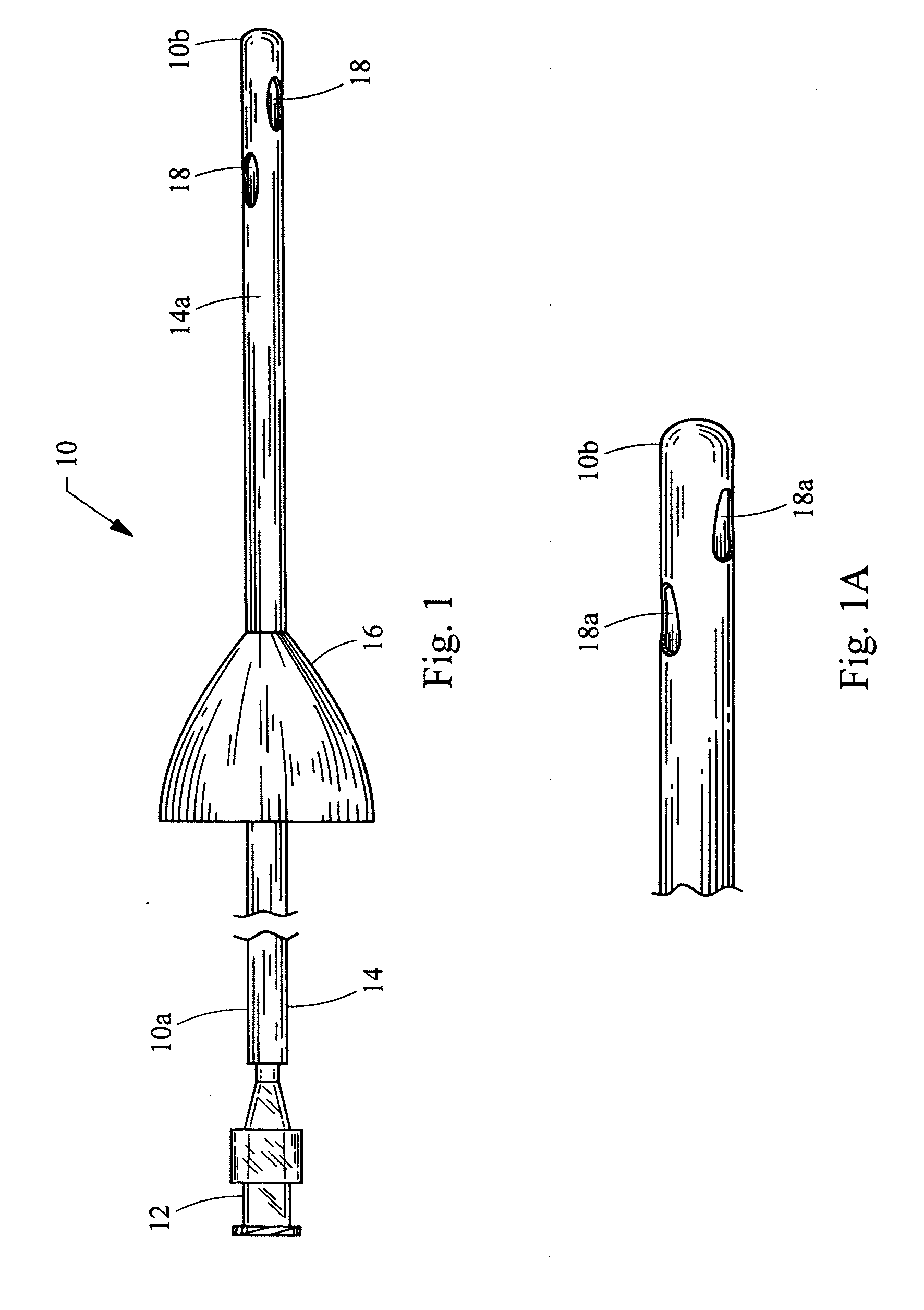
[0033] The device described below provides a way to occlude the cervix, to deliver and remove an image enhancing fluid into and from the uterus, and to take a biopsy of the endometrium. Embodiments of the device provide an effective and safe procedure for performing a sonohysterography and an endometrial biopsy. The embodiments are particularly useful for diagnosing abnormal uterine bleeding, such as that associated with hyperplasia. The embodiments are not limited for use with a human.

Problems solved by technology

The use of multiple devices to perform sonohysterography and endometrial biopsy may increase cramping of the uterus, patient pain, and patient discomfort.
The use of multiple devices may also increase the amount of time necessary to complete the procedures, increase patient recovery time, increase the risk of injury to the patient, and may involve greater expense.

Method used


Image

  • Sonohysterography and biopsy catheter
  • Sonohysterography and biopsy catheter
  • Sonohysterography and biopsy catheter

Examples


Embodiment Construction

[0033] The device described below provides a way to occlude the cervix, to deliver and remove an image enhancing fluid into and from the uterus, and to take a biopsy of the endometrium. Embodiments of the device provide an effective and safe procedure for performing a sonohysterography and an endometrial biopsy. The embodiments are particularly useful for diagnosing abnormal uterine bleeding, such as that associated with hyperplasia. The embodiments are not limited for use with a human.

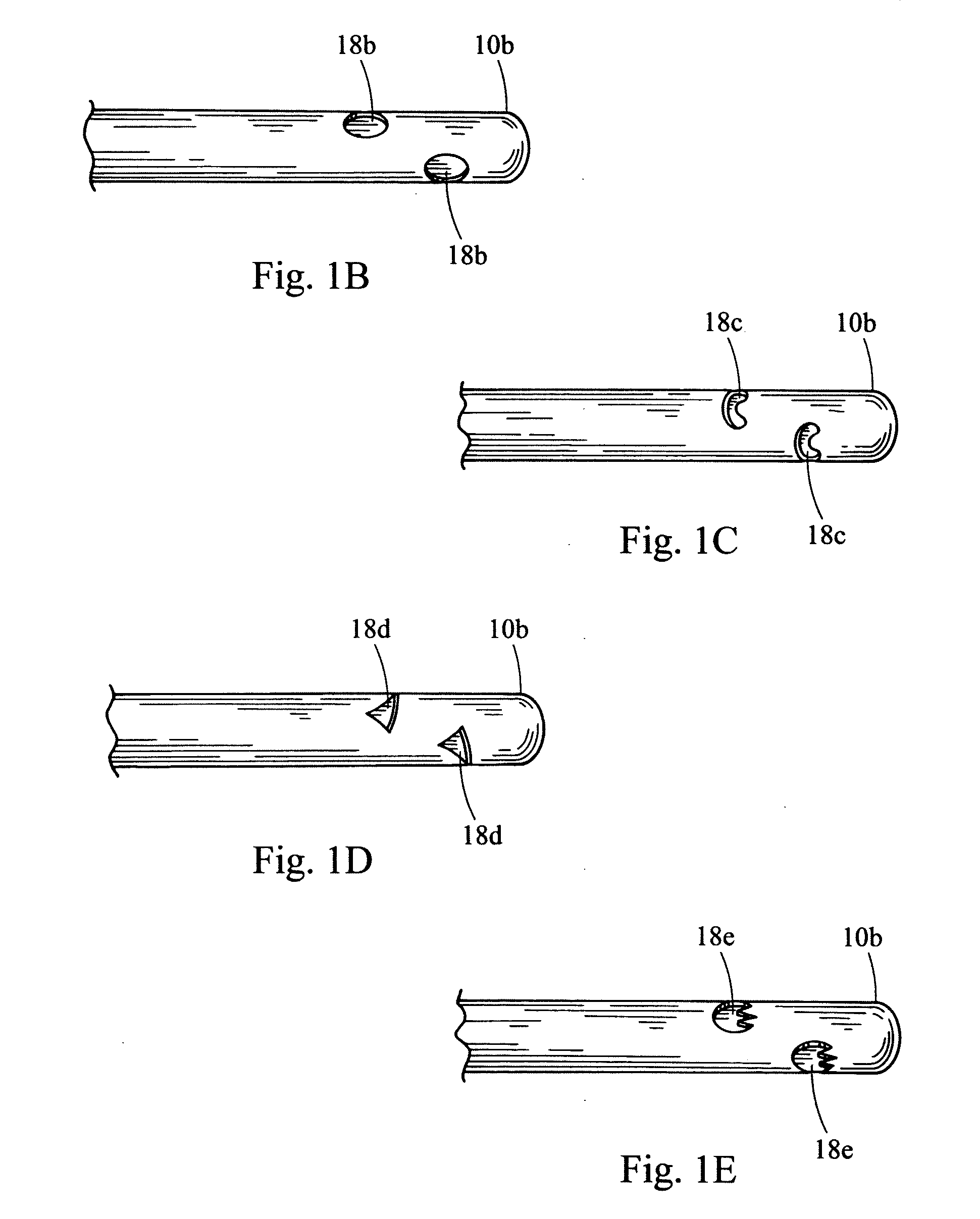
[0034] A more detailed description of the embodiments will now be given with reference to FIGS. 1-14. The present invention is not limited to those embodiments illustrated; it specifically contemplates other embodiments not illustrated and described but intended to be included in the claims. FIG. 1 is a plan view of a first embodiment of the device. A catheter assembly 10 has a proximal portion 10a, a distal portion 10b, and an elongated tubular body 14 having a lumen 14a extending throughout. Locate...


PUM


Abstract

A single device is provided for performing the diagnostic procedures of sonohysterography and endometrial biopsy. The device includes a cervical seal to occlude the cervix and a substance delivery and removal device to deliver and remove a fluid from the uterus. The substance delivery and removal device may also be used to create a vacuum so that suction occurs near side-ports that are used to collect endometrial tissue samples from multiple locations as the device is rotated or moved laterally within the patient. Additionally, the sample may be taken using a cellular collection device.

Description

RELATED APPLICATIONS [0001] The present patent document claims the benefit of the filing date under 35 U.S.C. §119(e) of Provisional U.S. Patent Application Ser. No. 60 / 787,760, filed Mar. 31, 2006, which is hereby incorporated by reference in its entirety.FIELD OF THE INVENTION [0002] This invention relates to the diagnosis of uterine abnormalities, such as abnormal uterine bleeding and for performing diagnostic procedures for a uterus. BACKGROUND OF THE INVENTION [0003] Medical professionals often use two procedures in order to diagnose abnormal uterine bleeding. The first procedure is sonohysterography, a technique employed for imaging the uterine cavity. Sonohysterography is commonly performed using a Goldstein catheter which is described in U.S. Pat. No. 6,706,026, assigned to the assignee of the present invention, and which is hereby incorporated in its entirety. Sonohysterography is performed by threading a catheter transcervically into the uterine cavity and delivering salin...

Claims


Application Information

Patent Timeline
Patent Type & Authority Applications(United States)
IPC IPC(8): A61M31/00
CPCA61B10/0045A61B17/42A61B2017/3419A61B2010/0216A61B2010/0074
Inventor COX, TED R.SHWAYDER, JAMES M.BILTZ, BENJAMIN T.WARD, JOHN H.WHETHAM, DANIEL K.WINGLER, TROY W.
Owner VANCE PROD INC D B A COOK UROLOGICAL INC
Learn More