Method and device for separate three-dimensional presentation of arteries and veins in a part of body

a three-dimensional presentation and artery technology, applied in the field of three-dimensional presentation of arteries and/or veins in a part of the body, can solve the problem of not being able to make a sufficiently clear distinction between arterial and venous vessels

Inactive Publication Date: 2007-10-04
SIEMENS HEALTHCARE GMBH
View PDF3 Cites 13 Cited by
  • Summary
  • Abstract
  • Description
  • Claims
  • Application Information

AI Technical Summary

Benefits of technology

[0009]The idea underlying the invention is to execute more than one filling run and to stagger these in time in relation to the contrast injection so that all parts of the imaged part of the body are imaged at least once in the arterial phase of the vessel contrasting to make a three-dimensional reconstruction possible.

Problems solved by technology

However, with conventional 3D angiography it is not possible to make a sufficiently clear distinction between arterial and venous vessel system because of the recording times and the dynamics of the contrast medium propagation.

Method used


Image

  • Method and device for separate three-dimensional presentation of arteries and veins in a part of body
  • Method and device for separate three-dimensional presentation of arteries and veins in a part of body
  • Method and device for separate three-dimensional presentation of arteries and veins in a part of body

Examples


Embodiment Construction

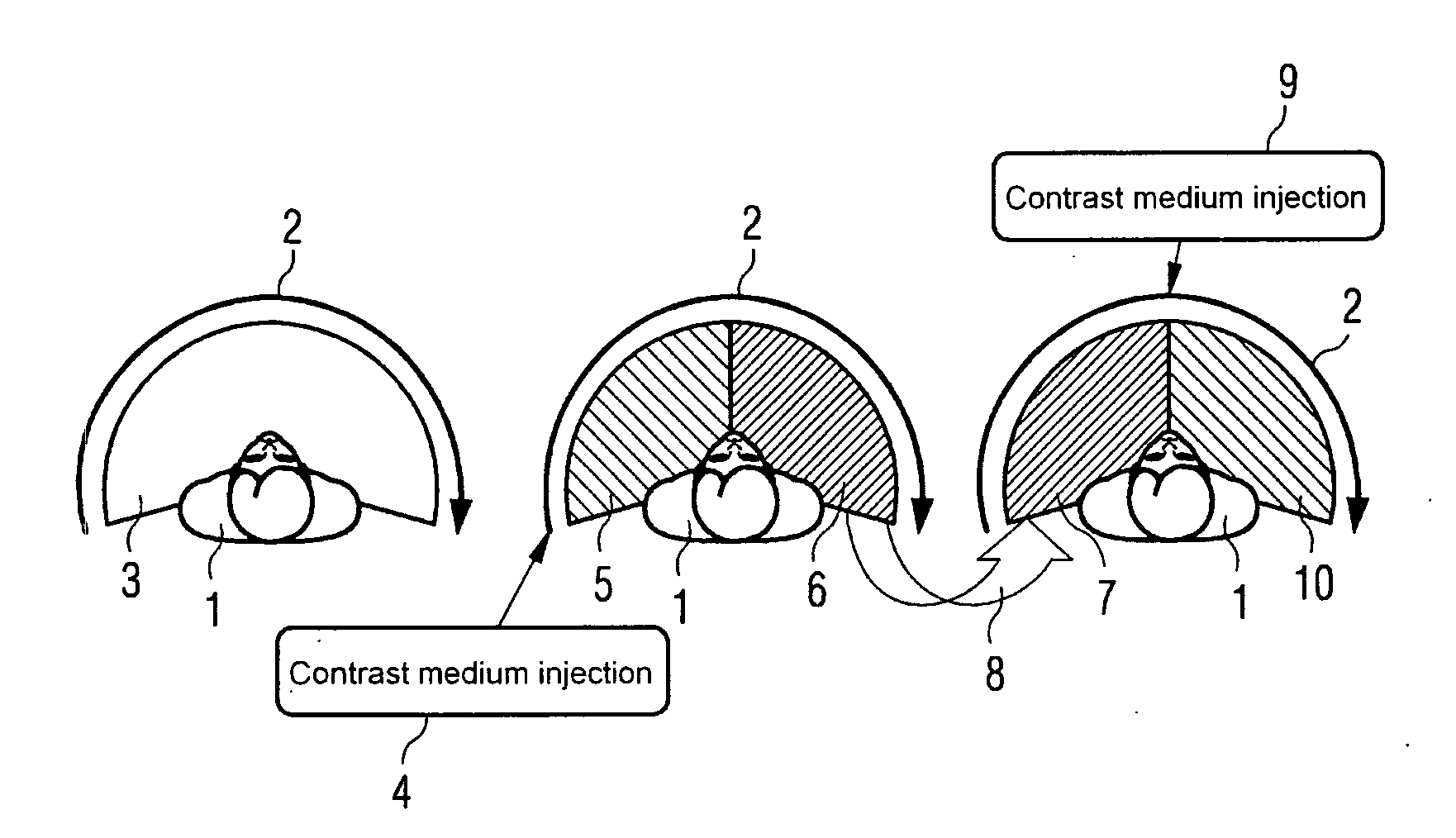
[0041]FIG. 1 describes the inventive method when using a C-arm angiograph which can only perform scanning runs in one direction.

[0042]The left-hand part of FIG. 1 shows a body positioned for examination, such as a patient 1, in cross-section, arrow 2 shows the direction of scanning of the C-arm angiograph used and sector 3 shows the detected projection angle of the masking run. Here the entire part of the body is recorded and represented in accordance with the options provided by the angiograph used. The middle part of FIG. 1 shows the first filling run, in which, at the beginning of the second scanning run in the same scanning direction 2, a contrast medium is injected into or adjacent to the vessel to be examined. FIG. 1 illustrates the point in time of the contrast medium injection 4. The cross-hatched area 5 is the area of the filling run, in which the arterial phase of the vessel contrasting occurs. After the end of this phase 5, during which the contrast medium has reached the...


PUM


Abstract

The invention is directed to a method and a device for separate three-dimensional presentation of arteries and/or veins of a vessel system in a part of the body of a vertebrate by a rotation computer tomography. A masking run of the tomograph is undertaken without contrast media around the part of the body. Then two filling runs with contrast media are executed, with the venous phase of vessel contrasting during the first filling run occurring during the arterial phase of vessel contrasting of the second filling run and vice-versa. The data from the first and second filling run is combined into data sets from the arterial or the venous phases of the vessel contrasting of the filling runs. The data of the masking run is subtracted from the combined data sets to obtain final data sets for a three-dimensional presentation of the arterial or the venous vessel system.

Description

CROSS REFERENCE TO RELATED APPLICATIONS[0001]This application claims priority of German application No. 10 2006 012 181.3 filed Mar. 16, 2006, which is incorporated by reference herein in its entirety.FIELD OF THE INVENTION[0002]The present invention relates to a method for separate three-dimensional presentation of arteries and / or veins of a vessel system in a part of the body of a vertebrate by means of a rotation computer tomograph as well as a device, especially for execution of the method. Preferred applications of the invention are for C-arm angiographs.BACKGROUND OF THE INVENTION[0003]There is a growing demand for the most accurate possible three-dimensional presentation of the appearance and course of vessels in parts of the body, especially of arteries and veins, for diagnostic purposes in the field of vessel diseases and their treatment. The examination of cerebral aneurysms represents an important area of application, which also includes an analysis and optimum presentati...

Claims


Application Information

Patent Timeline
Patent Type & Authority Applications(United States)
IPC IPC(8): A61B5/05A61B6/00A61B6/03A61B8/06
CPCA61B6/504A61B6/481
Inventor BENNDORF, GOETZKLINGENBECK-REGN, KLAUSZELLERHOFF, MICHAEL
Owner SIEMENS HEALTHCARE GMBH
Why Eureka
Try Eureka