Thorax mounted stabilization platform

a technology of a stabilization platform and a thorax, which is applied in the field of abdominal and thoracic surgical devices, can solve the problems of significant physical trauma to the patient, requiring one week of hospital recovery time, and weeks of convalescen

Inactive Publication Date: 2005-05-12
ESTECH ENDOSCOPIC TECH
View PDF26 Cites 4 Cited by
  • Summary
  • Abstract
  • Description
  • Claims
  • Application Information

AI Technical Summary

Benefits of technology

[0010] In keeping with the foregoing discussion, the present invention provides a thorax mounted stabilizing platform for a surgical device, such as a tissue stabilizer, that can be inserted through a separate minimally invasive incision and that does not need to be mounted on the surgical retractor or the surgical table for stability. The stabilizing platform can be affixed to the thorax of a patient during a surgical procedure. A rod is introduced into a percutaneous opening in the patient. An internal and / or an external fixing device is deployed to attach the rod to the patient. One or more surgical devices may be mounted to the distal or internal end of the rod. An adjustment knob or other actuation mechanism is located at the proximal or external end of the rod to actuate or manipulate the surgical device(s) attached to the distal end.

Problems solved by technology

This technique causes significant physical trauma to the patient and can require one week of hospital recovery time and up to eight weeks of convalescence.
This can be very expensive in terms of hospital costs and disability, to say nothing of the pain to the patient.
While this procedure has the advantage of being less invasive or traumatic than performing a media, sternotomy, there are numerous disadvantages to using trocars to establish the entry ports for the instruments and viewscope.
In addition, because of the limited movement, the surgeon often has to force the instrument into an angle that moves the trocar and further damages the surrounding tissue and nerves.
The need to force the instrument causes the surgeon to lose sensitivity and tactile feedback, thus making the surgery more difficult.
A disadvantage of this approach is that the tissue stabilizer tends to crowd the surgical field, which is particularly a problem when using small minimally invasive incisions for performing the surgery.

Method used


Image

  • Thorax mounted stabilization platform
  • Thorax mounted stabilization platform
  • Thorax mounted stabilization platform

Examples


first embodiment

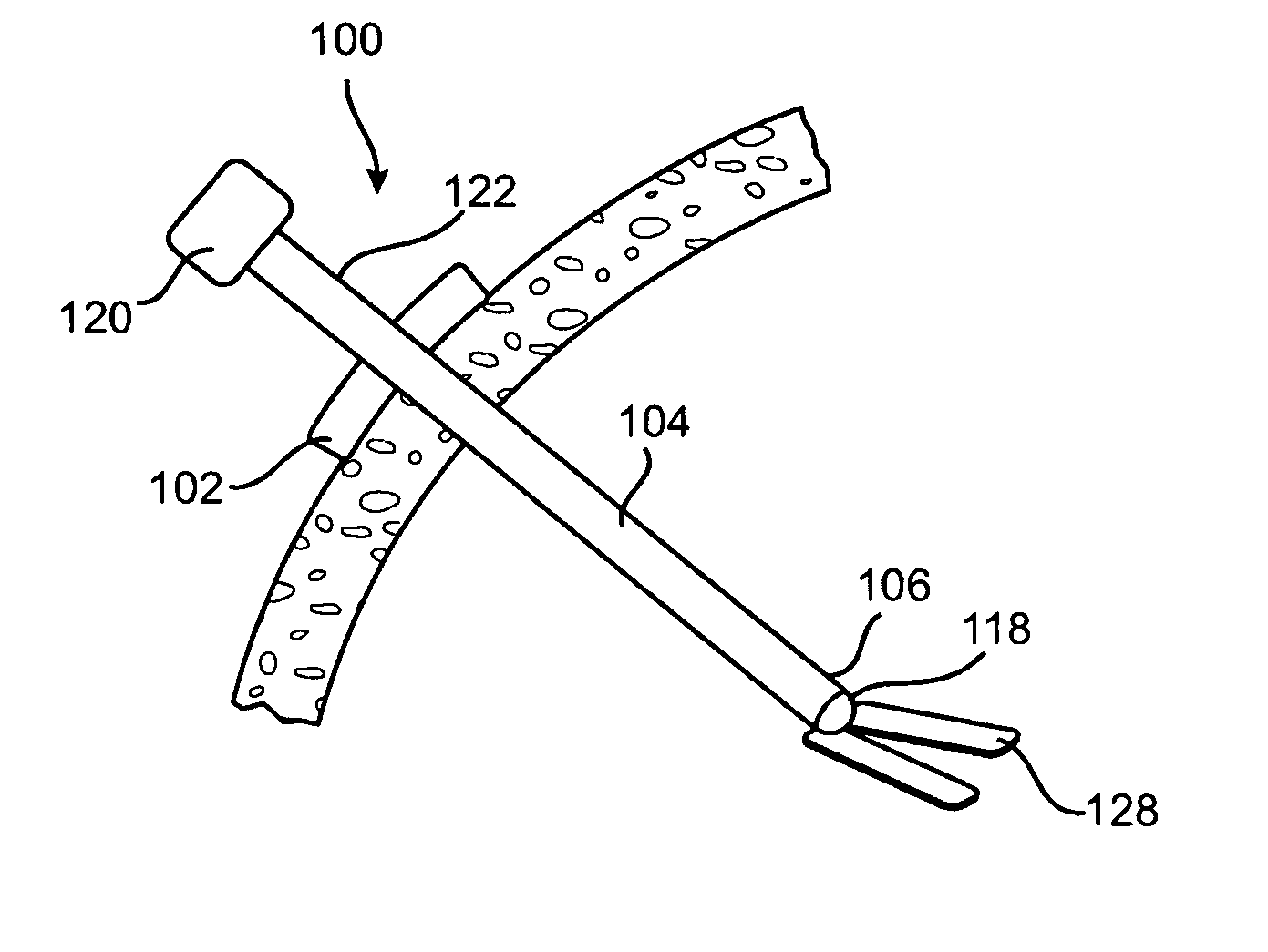
[0016]FIG. 1 shows the stabilization platform 100. In this embodiment, an external stabilizer 102 is used to hold the platform in place during the surgical procedure being performed. The external stabilizer 102 is an object located around or attached to a rod 104 that prevents the distal end 106 of the rod from extending too far into the patient. The external stabilizer 102 may be located at a fixed point on the rod 104. Alternatively, the user may select a depth to which the end of the surgical instrument or rod 104 should extend and then move the external stabilizer 102 to the appropriate location along the rod 104. The rod 104 may have depth markers to assist in gauging the appropriate depth. Alternatively, the user may guide the distal end 106 of the rod 104 and / or surgical tool into place by feel or using a known imaging system. Then, the user would slide or place the external stabilizer 102 and affix it to the selected location on the rod 104. To further secure the external st...

second embodiment

[0017]FIG. 2 shows the stabilization platform. In this embodiment, an internal stabilizer 110 is used to hold the device in place during the surgical procedure being performed. The internal stabilizer 110 is an object located around or attached to the rod 104 that prevents the device from being inadvertently removed from the patient or may be used to seal the opening through the wall of the cavity in the patient.

[0018] The internal stabilizer 110 may take the form of an elongated member. The narrow direction of the internal stabilizer is sized to fit between the ribs of the patient. Once the internal stabilizer 110 is inserted into the patient, the internal stabilizer is rotated 90 degrees. The long direction of the internal stabilizer 110 is sized such that, after rotation, the ends of the elongated member 110 rest against the internal surface of the thoracic cavity.

[0019] In another version, the internal stabilizer is inflatable. Once the internal stabilizer has passed through th...

third embodiment

[0022]FIG. 3 shows the stabilization device 100. In this embodiment, both an external stabilizer 102, as described in FIG. 1, and an internal stabilizer 110, as described in FIG. 2, are used to hold the device in place during the surgical procedure being performed. With the use of both the internal and external stabilizers, the device is locked into place and cannot penetrate farther into the patient or move back out of the patient. In this version, the internal stabilizer and external stabilizer may be formed of one or more projections, which act as a clamp and may be selectively placed around a stable structure in the patient, such as a rib. If desired, two pair of projections may be used. In this case, the two pair can clamp onto two adjacent ribs. Additional pairs of projections may be used to further secure the device.

[0023] Alternate versions of the above embodiments may be configured with internal or external clamps to attach to other surgical tools, such as the retractor use...


PUM


Abstract

A thorax mounted stabilizing platform for a surgical device, such as a tissue stabilizer, can be inserted through a minimally invasive incision and affixed to the thoracic wall to stabilize the surgical device. The stabilizing platform includes a rod that is introduced into a percutaneous opening in the patient. An internal and/or an external fixing device is deployed to attach the rod to the patient. One or more surgical devices may be mounted to the distal or internal end of the rod. An adjustment knob or other actuation mechanism is located at the proximal or external end of the rod to actuate or manipulate the surgical device(s) attached to the distal end.

Description

CROSS REFERENCE TO RELATED APPLICATIONS [0001] This application claims the benefit of U.S. Provisional Application No. 60 / 519,221, filed on Nov. 11, 2003. This and all patents and patent applications referred to herein are hereby incorporated by reference in their entirety.FIELD OF THE INVENTION [0002] The invention pertains to apparatus and methods for delivery and use of surgical devices. In particular, it is a stabilization platform mounted on the thorax of the patient for use with endoscopic surgical tools. BACKGROUND OF THE INVENTION [0003] Surgery on the heart is one of the most commonly performed types of surgery that is done in hospitals across the U.S. Cardiac surgery can involve the correction of defects in the valves of the heart, defects to the veins or the arteries of the heart and defects such as aneurysms and thromboses that relate to the circulation of blood from the heart to the body. Coronary artery bypass graft (CABG) surgery is one of the most common cardiac surg...

Claims


Application Information

Patent Timeline
Patent Type & Authority Applications(United States)
IPC IPC(8): A61B1/32A61B17/00A61B17/02A61B17/34
CPCA61B17/34A61B2017/00243A61B2017/0243A61B2017/3492A61B2017/3484A61B2017/3486A61B2017/3482
Inventor BERTOLERO, ARTHUR A.IBRAHIM, TAMER
Owner ESTECH ENDOSCOPIC TECH
Why Eureka
Try Eureka