
Photochemotherapeutic method using 5-aminolevulinic acid and other precursors of endogenous porphyrins

a technology of endogenous porphyrin and photochemotherapy, which is applied in the direction of biocide, heterocyclic compound active ingredients, peptide/protein ingredients, etc., can solve the problems of inconvenient long time, inconvenient exposure, and persisting clinically significant porphyrin amount,

Inactive Publication Date: 2004-06-10
QUEENS UNIV OF KINGSTON
View PDF20 Cites 2 Cited by
  • Summary
  • Abstract
  • Description
  • Claims
  • Application Information

AI Technical Summary

Benefits of technology

0018] This invention is based on the finding that exogenously administered ALA and other precursors of PpIX are metabolized in patients to PpIX and that PpIX preferentially accumulates in rapidly growing cells, as contrasted with less rapidly growing cells. The rapid growth is correlated with the metabolic activity, so that the differential accumulation is affected by the relative metabolic activity between different cells.
0019] This invention provides a method for detecting in a patient, a malignant or non-malignant lesion or abnormality which is sensitive to PpIX, namely those which preferentially accumulate PpIX, comprising administering to said patient an effective amount of a precursor of PpIX in the biosynthetic pathway for heme so as to induce an accumulation of PpIX in said lesions, and exposing said lesions to light having a wavelength within the absorption spectrum of said PpIX, thereby to induce fluorescence in said lesions.
0020] Another aspect of this invention is a method for treating malignant and non-malignant hyperproliferative lesions of the skin, mucosa, endometrium and urothelium which are sensitive to PpIX in a patient, comprising administering to said patient an effective amount of a precursor of PpIX in the biosynthetic pathway for heme so as to induce synthesis or accumulation or both of PpIX or other endogenous porphyrins, their precursors and their photoproducts in said lesions, and exposing said lesions to light having a wavelength within the photoactivating action spectrum of said PpIX to thereby induce photoactivation in said lesions.
0021] Thus, the rapidly growing cells involved can be either malignant or non-malignant hyperproliferative cells. The hyperproliferative cells can be normal, rapidly growing cells or abnormal cells in otherwise normal tissue. The abnormal cells in an otherwise normal tissue can include abnormal rapidly growing cells endogenous to the patient or abnormal, rapidly growing cells which are exogenous to the patient. These rapidly growing cells that are exogenous to the patient shall, for convenience, be referred to hereby, depending on the degree of generality, as rapidly growing exogenous cells, rapidly growing Protista cells and rapidly growing parasite cells.
0022] One aspect of this invention is induction in vivo or in vitro of the biosynthesis and selective accumulation of fluorescing or photosensitizing concentrations of protoporphyrin IX or other endogenous porphyrins such as coproporphyrin I, coproporphyrin III, uroporphyrin I, uroporphyrin III, or fluorescent metalloporphrins such as zinc protoporphyrin IX in Protista and parasites of humans or other animals, by exposing said Protista and endogenous cells under appropriate conditions in vivo or in vitro to an effective concentration of 5-aminolevulinic acid or other precursor of said porphryin(s) in the biosynthetic pathway for heme.

Problems solved by technology

Unfortunately, a clinically significant (photosensitizing) amount of porphyrin may persist in the skin for at least two weeks, (occasionally for more than two months) following the intravenous injection of HpIX, HpD, or a semi-puridied preparation of HpD, such as Photofrin II.
This means that patients must avoid exposure to sunlight (either direct, or through window glass) for an inconveniently long period of time post-injection.
Understandably, patient compliance often is poor, and accidental phototoxic "sunburn" is a common occurrence in the weeks following a diagnostic or therapeutic injection of porphyrin.
Persistent photosensitivity is the major hazard associated with this technique, and is the main reason why it is not used more widely.
Localized exposure of psoralen-containing tissues to ultraviolet light induces a localized photochemical reaction that causes the drug to bind covalently to the DNA of living cells, thus destroying their proliferative potential.
However, there are two serious problems with such treatments.
First, the procedure has been demonstrated in humans to be carcinogenic.
Second, the depth at which malignant tissue can be killed is limited to a few millimeters below the illuminated surface.
These problems severely limit the usefulness of the methoxypsoralens for photochemotherapy.
When porphyrins are used as photosensitizers, cell death results from damage to cell membranes.
The main problem with the systemic use of HpIX, HpD and Photofrin II is that photosensitizing concentrations persist in the skin for several weeks to several months following their administration.
Consequently, severe accidental phototoxic skin reactions may occur unless the patient avoids exposure to sunlight (either direct, or filtered through window glass) until the concentration of the photosensitizer in the skin has been reduced to a harmless level.
Not all patients comply with these instructions, since it often is quite inconvenient to do so.
However, another type of problem is encountered if HpIX or HpD is applied topically in DMSO (dimethylsulfoxide), Azone, or some other vehicle intended to enhance their diffusion through tissue.
Consequently, the topical application of porphyrins often is associated with a loss of specificity for malignant tissues, and normal tissues near the site of application may develop persistent photosensitization from the localized concentration of porphyrin.

Method used


Image

  • Photochemotherapeutic method using 5-aminolevulinic acid and other precursors of endogenous porphyrins
  • Photochemotherapeutic method using 5-aminolevulinic acid and other precursors of endogenous porphyrins

Examples


example 1

[0065] Long Term Photodynamic Endometrial Ablation--Rats were divided into 2 groups (6 and 7 rats / group) and their uterine horns were injected with 4 or 8 mg ALA. Example 1, of U.S. application Ser. No. 08 / 082,113, filed Jun. 21, 1993 (U.S. Pat. No. 5,422,093), was repeated with the exception that all rats were exposed to light and the time from ALA administration to breeding was extended from 10-20 days to 60-70 days. All other procedures were identical to Example 1.

[0066] Breeding 60-70 days after photodynamic treatment with 4 mg ALA resulted in no implantations in the uterine horns treated with ALA (n=6) whereas fetuses were found in all control uterine horns treated with saline (n=6). These results confirmed the long term endometrial ablative effect of PDT. In the groups of rats (n=7) treated with 8 mg ALA 2 of 7 became pregnant in ALA treated uterine horns compared with 7 of 7 pregnancies in the saline treated horns.

[0067] Histology--In order to show normal uterine histology of...

example 2

[0068] The procedures of Example 1 (U.S. Pat. No. 5,422,093) were repeated with 1, 2, 3, 4 and 5 hour incubation periods using a level of 1 mM of ALA. No significant fluorescence was observed in the myometrial samples or in the endometrial samples incubated for 2 hours. Maximum fluorescence was observed in the endometrial samples incubated for 4 hours.

example 3

[0069] Endometrial Fluorescence in vivo following Topical Application of ALA in the Non-human Primate--50 mg of ALA was injected into the uterine lumen of an adult, healthy, female rhesus monkey following exposure of the uterus at laparotomy. A hysterectomy was performed 3 hours later and cross sectional slices incorporating endometrial and myometrial tissue were taken from the uterine specimen. These slices were subjected to examination by fluorescence microscopy as in Example 2 and 3 above. Fluorescence was observed throughout the endometrium of all slices. No fluorescence was observed in the myometrium.

[0070] The above examples clearly illustrate that endometrial ablation in a range of animal species, including humans, by photodynamic therapy using ALA can be achieved with little or no damage to the underlying myometrial tissues.


PUM

PropertyMeasurementUnit
wavelengthaaaaaaaaaa

Abstract

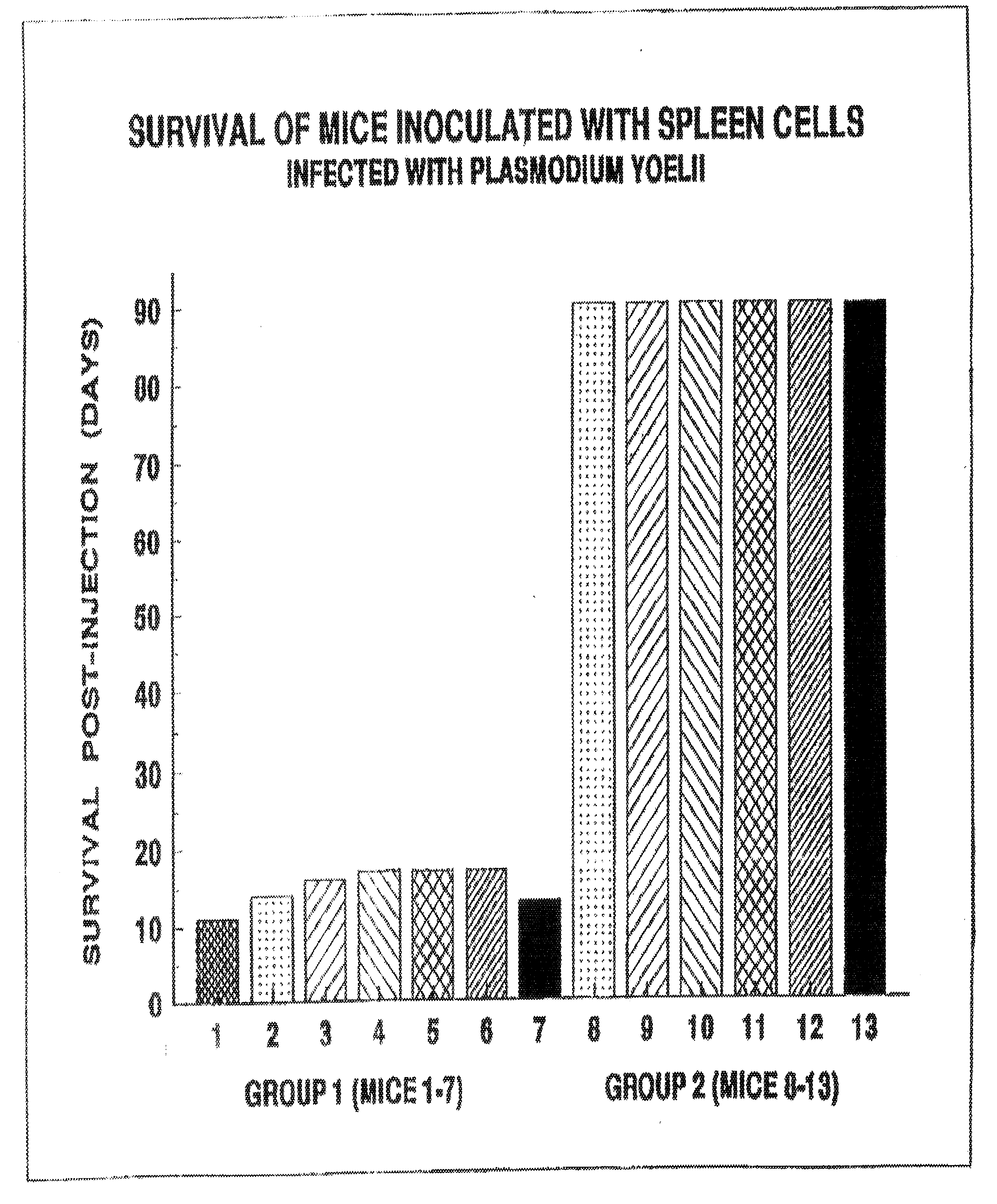
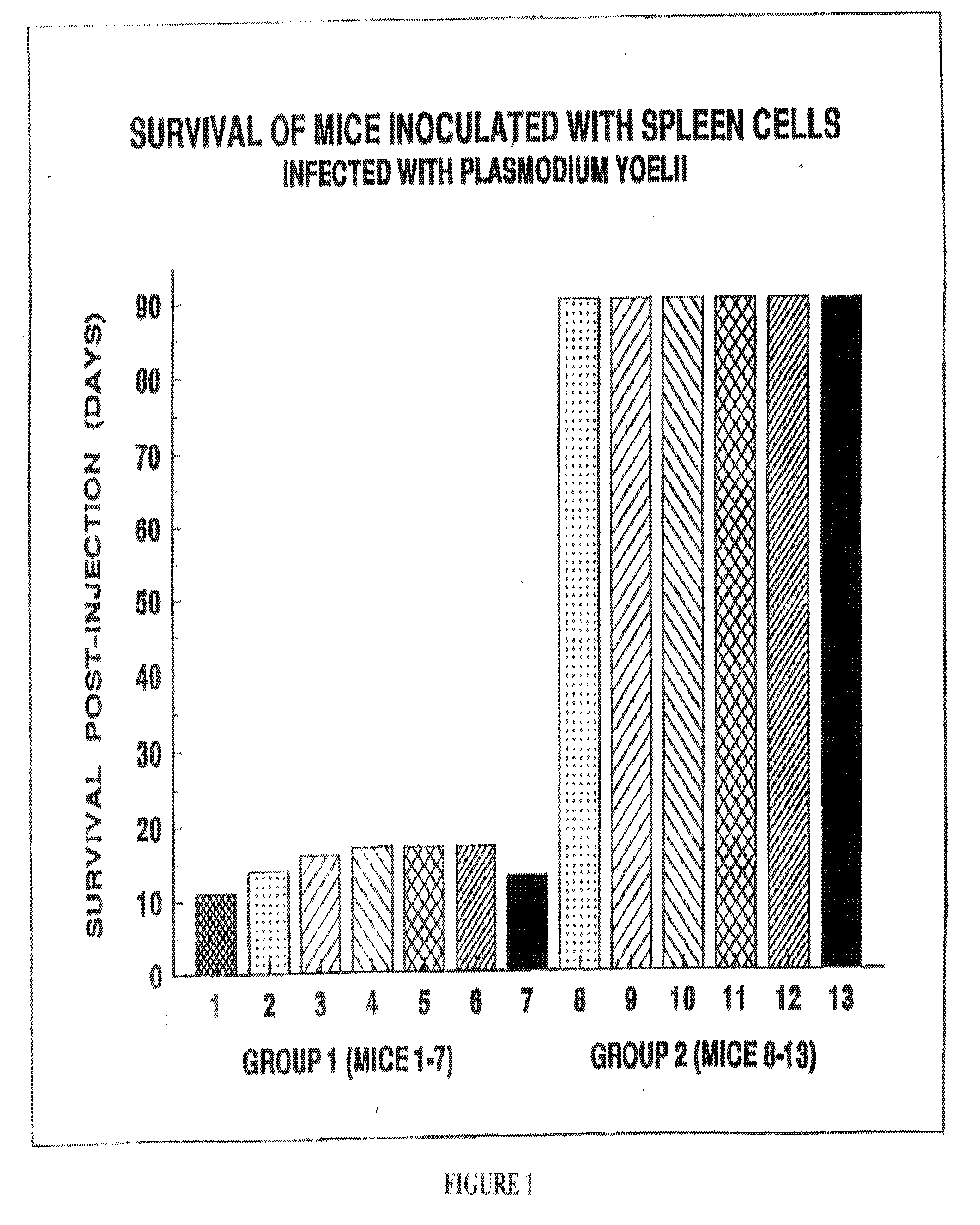
Methods of detecting and treating rapidly growing exogenous cells, such as Protista, or parasites, that preferentially accumulate a photoactivatable porphyrin in which 5-aminolevulinic acid or precursor thereof is administered to the patient, or contacted to the exogenous cells, in an amount sufficient to induce synthesis fluorescence and / or photosensitizing concentrations of a protoporphyrin IX in the exogenous cells, followed by exposure of the exogenous cells to light of photoactivating wavelengths.

Description

[0001] This is a Divisional Application of application Ser. No. 09 / 816,329, filed Mar. 26, 2001, which is a Continuation Application of application Ser. No. 09 / 293,835, filed Apr. 19, 1999, which is in turn a Continuation-in-part of application Ser. No. 08 / 082,113 (now U.S. Pat. No. 5,422,093), filed Jun. 28, 1993, which is in turn a Continuation-in-part of application Ser. No. 07 / 865,151 (now U.S. Pat. No. 5,234,940), filed Apr. 8, 1992, which is in turn a Continuation-in-part of application Ser. No. 07 / 783,750 (now U.S. Pat. No. 5,211,938), filed Oct. 28, 1991, which is in turn a Continuation of application Ser. No. 07 / 386,414 (now U.S. Pat. No. 5,079,262), filed Jul. 28, 1989.[0002] This invention relates to the detection and treatment, by induced fluorescence and photochemotherapy, respectively, of certain tissue abnormalities (both cancerous and non-malignant of endogenous and exogenous origin), hyperproliferative cells, and normal cells. The invention also relates to the detec...

Claims


Application Information

Patent Timeline
Patent Type & Authority Applications(United States)
IPC IPC(8): A61K31/195A61K31/555A61K41/00
CPCA61K31/195A61K41/0061A61K31/555
Inventor KENNEDY, JAMES C.POTTIER, ROY H.
Owner QUEENS UNIV OF KINGSTON
Learn More