Energy-sensitive multi-contrast cost-effective CT system

a multi-contrast, cost-effective technology, applied in the field of energy-sensitive multi-contrast cost-effective ct system, can solve the problems of ct and other types of ct imaging, x-rays lack the power to observe biological soft tissues, etc., and achieve the effects of small size, fast data acquisition, and convenient us

Active Publication Date: 2022-03-08
RENESSELAER POLYTECHNIC INST
View PDF14 Cites 2 Cited by
  • Summary
  • Abstract
  • Description
  • Claims
  • Application Information

AI Technical Summary

Benefits of technology

[0005]Embodiments of the subject invention provide novel and advantageous systems and methods for obtaining scattering images (e.g., small angle scattering images or dark field images) during computed tomography (CT) imaging (e.g., X-ray CT imaging). Two gratings or grating layers can be disposed between the object to be imaged and the detector, and the gratings or grating layers can be arranged such that primary X-rays (those that are not scattered, or deflected as they pass through the object to be imaged) are blocked (e.g., partially, mostly, or completely blocked) while scattered X-rays (e.g., small angle scattering X-rays) that are deflected as they pass through the object to be imaged reach the detector to generate the scattering image (e.g., small angle scattering image or dark field image). This can lead to high-resolution, accurate dark field images that can have many important applications, including identification of cancer tissue in an animal, such as a mammal (e.g., a human). Compared to related art imaging systems and methods, systems and methods of embodiments of the subject invention are relatively efficient, inexpensive, easy to use, fast in data acquisition, and / or small in size. Energy-sensitive, multi-contrast cost-effective CT systems are provided herein, and can be termed EMCC or E=MC2 systems for short.

Problems solved by technology

The X-ray absorption coefficient is roughly proportional to the fourth power of the atomic number, and that is why X-rays lack sufficient power to observe biological soft tissues.
Existing techniques for micro CT and other types of CT imaging all have drawbacks, though.

Method used


Image

  • Energy-sensitive multi-contrast cost-effective CT system
  • Energy-sensitive multi-contrast cost-effective CT system
  • Energy-sensitive multi-contrast cost-effective CT system

Examples


example 1

[0058]EGSnrc simulation software was used to simulate small angle scattering imaging using a system according to an embodiment of the subject invention. Monte Carlo simulations were performed on the system.

[0059]EGSnrc is a well-known simulation software whose function is to model the passage of electrons and photons through matter. Because it relies on Monte Carlo, which is the most accurate method to model the transport of radiation, the simulation results discussed herein are logical and reliable.

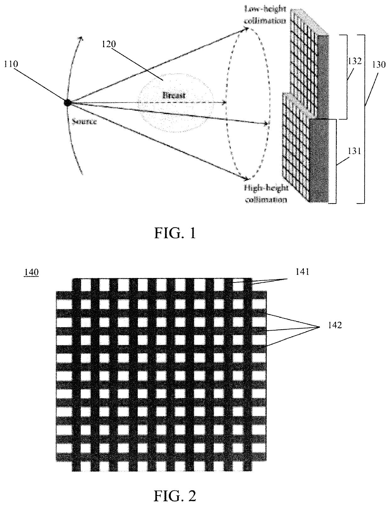
[0060]The gratings were composed of tungsten. The height and width of each grating cell were set to be 1 centimeter (cm) and 0.5 millimeter (mm), respectively. The dimensions of the detector were 100 mm×3 mm with a pixel size of 1 mm. The grating was aligned with the middle of the detector. The length of the gap between the two layers of the grating was 0.1 cm. The X-ray source was a parallel-beam source with an operating voltage of 20 kilovolts-peak (kVp), and there was a total of 1.28×...

example 2

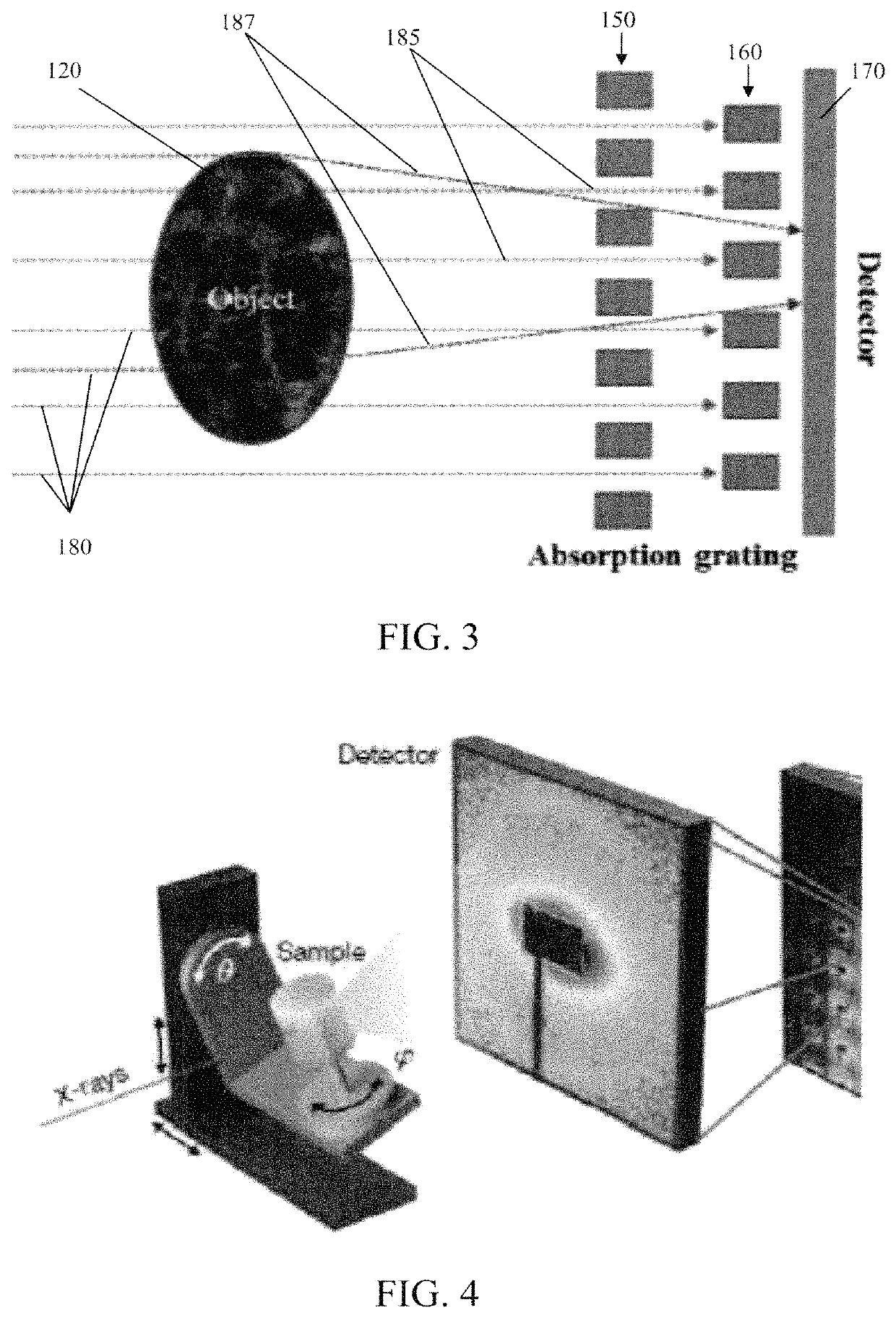
[0063]A second simulation experiment was performed using the same parameters as in Example 1, but only one phantom was used. The phantom 121 was located in front of the middle of the grating 150. In order to reconstruct the scattering data, the phantom was rotated 360 degrees equiangularly around its center, and 10 groups of scattering data were acquired. The profile of the average value of these 10 groups of scattering data is shown in FIG. 10 (the line that is less smooth). It can be seen that the quadratic fit curve (the smoother line) fits well with the profile, and that means the scattering signal is effective. By the physical model to describe X-ray small angle scattering based on the principle of energy conservation, these ten groups of scattering data were used to reconstruct the cross-sectional image of the phantom 121, which is shown in FIG. 11. The shape and size of the circle in FIG. 11 is completely consistent with the phantom. This result demonstrates that the scatteri...


PUM


Abstract

Systems and methods for obtaining scattering images during computed tomography (CT) imaging are provided. Two gratings or grating layers can be disposed between the object to be imaged and the detector, and the gratings or grating layers can be arranged such that primary X-rays are blocked while scattered X-rays that are deflected as they pass through the object to be imaged reach the detector to generate the scattering image.

Description

CROSS-REFERENCE TO RELATED APPLICATION[0001]This application is a national stage application of International Patent Application No. PCT / US2017 / 018456, filed Feb. 17, 2017, which claims the benefit of U.S. Provisional Application Ser. No. 62 / 296,204, filed Feb. 17, 2016, which is incorporated herein by reference in its entirety, including any figures, tables, and drawings.STATEMENT REGARDING FEDERALLY SPONSORED RESEARCH OR DEVELOPMENT[0002]This invention was made with government support under grant No. EB016977 awarded by the National Institutes of Health. The government has certain rights in the invention.BACKGROUND[0003]High-resolution X-ray assessment of lung and other tumors, in vivo or after surgical resection, has become increasingly important, as are imaging studies of small objects such as organoids. Samples, specimens, and tissue constructs have several unique properties, including but not limited to: less issues with increased radiation dose given that they are samples ins...

Claims


Application Information

Patent Timeline
Patent Type & Authority Patents(United States)
IPC IPC(8): A61B6/00A61B6/04A61B6/03
CPCA61B6/4291A61B6/03A61B6/0407A61B6/483A61B6/484A61B6/508A61B6/032
Inventor WANG, GECONG, WENXIANG
Owner RENESSELAER POLYTECHNIC INST
Why Eureka
Try Eureka