Ultrasonic imaging method and system

An ultrasonic imaging method, ultrasonic technology, applied in the direction of ultrasonic/sonic/infrasonic Permian technology, ultrasonic/sonic/infrasonic image/data processing, ultrasonic/sonic/infrasonic diagnosis, etc., which can solve the problem of difficult maintenance of the target tissue inspection section Problems such as the same, difficult to display elastic images and contrast images at the same time

Pending Publication Date: 2021-05-11
SHENZHEN MINDRAY BIO MEDICAL ELECTRONICS CO LTD +1
View PDF0 Cites 2 Cited by
  • Summary
  • Abstract
  • Description
  • Claims
  • Application Information

AI Technical Summary

Problems solved by technology

On the one hand, it is difficult to maintain the same inspection section of the target tissue during successive operations; on the other hand, it is difficult to sim

Method used


Image

  • Ultrasonic imaging method and system
  • Ultrasonic imaging method and system
  • Ultrasonic imaging method and system

Examples


Embodiment Construction

[0213] The present invention will be further described in detail below through specific embodiments in conjunction with the accompanying drawings. Wherein, similar elements in different implementations adopt associated similar element numbers. In the following implementation manners, many details are described for better understanding of the present application. However, those skilled in the art can readily recognize that some of the features can be omitted in different situations, or can be replaced by other elements, materials, and methods. In some cases, some operations related to the application are not shown or described in the description, this is to avoid the core part of the application being overwhelmed by too many descriptions, and for those skilled in the art, it is necessary to describe these operations in detail Relevant operations are not necessary, and they can fully understand the relevant operations according to the description in the specification and genera...


PUM


Abstract

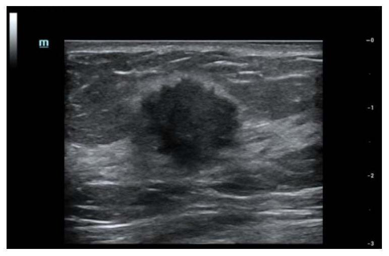
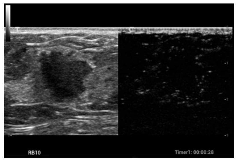
An ultrasonic imaging method comprises the following steps of emitting an ultrasonic wave to interested target tissue in a contrast imaging manner, and receiving an echo signal, to obtain contrast images of the interested target tissue within a period of time; determining a reference section in each contrast image, and meanwhile acquiring a tissue structure image corresponding to the reference section; emitting the ultrasonic wave to the interested target tissue to obtain current tissue structure images; matching the current tissue structure images with the tissue structure images corresponding to the reference sections, and determining to-be-examined section of current tissue structure images meeting the predetermined condition; emitting the ultrasonic wave to the interested target tissue in an elasticity imaging manner, to obtain an elasticity image of the interested target tissue on each to-be-examined section; and at least simultaneously displaying the contrast images of the reference sections and the elasticity images on the to-be-examined sections. Contrast imaging and elasticity imaging are carried out on a same section and the contrast images and the elasticity images are displayed at the same time, so that the operation process of a user is simplified, and the examination result is more accurate and reliable.

Description

technical field [0001] The present application relates to ultrasound imaging technology, in particular to a method and device for synchronously displaying ultrasound images in at least two imaging modes. Background technique [0002] Ultrasound elastography is one of the hot technologies in clinical research in recent years. It mainly uses ultrasound to detect the elasticity or softness and hardness of biological tissues, and outputs the elastic detection results. Restoration evaluation and other aspects have been more and more applied. [0003] Contrast-enhanced ultrasound imaging is an important inspection method in ultrasonic diagnosis. It mainly injects contrast agent into human blood vessels to enhance the Doppler signal of blood flow, so as to better observe the blood perfusion of tissues. It is used in clinical tumor detection. It plays an important role in diagnosis and auxiliary diagnosis. [0004] Among the existing commercial ultrasound products on the market, t...

Claims


Application Information

Patent Timeline
IPC IPC(8): A61B8/08A61B8/00
CPCA61B8/4416A61B8/4444A61B8/463A61B8/481A61B8/485A61B8/5246
Inventor 李双双周建桥
Owner SHENZHEN MINDRAY BIO MEDICAL ELECTRONICS CO LTD
Why Eureka
Try Eureka