
Surgical fixation device for fixing organs in minimally invasive surgery

A technology of minimally invasive surgery and fixation device, which is applied in the directions of stereotaxic surgical instruments, surgery, medical science, etc., can solve the problems of increasing the surgical space, large volume, inconvenient insertion into the minimally invasive incision, etc. Ease of surgery

Active Publication Date: 2021-03-19
THE SECOND AFFILIATED HOSPITAL TO NANCHANG UNIV
View PDF5 Cites 5 Cited by
  • Summary
  • Abstract
  • Description
  • Claims
  • Application Information

AI Technical Summary

Problems solved by technology

[0002] In minimally invasive surgery, surgeons often use large instruments to operate through small holes, and use endoscopic cameras to observe internal anatomical structures. Organs often interfere with the doctor's operation, so nurses are often required to use special surgical instruments to prop up, peel off, or fix these organs that have nothing to do with the lesion, so as to increase the operating space and facilitate the operation; However, the existing surgical instruments used to stretch or fix the organs are too large, which is inconvenient to insert into the minimally invasive incision. Excessively large surgical instruments will also cause the surgical wound to be too large, which is not conducive to postoperative recovery and aesthetics

Method used


Image

  • Surgical fixation device for fixing organs in minimally invasive surgery
  • Surgical fixation device for fixing organs in minimally invasive surgery
  • Surgical fixation device for fixing organs in minimally invasive surgery

Examples


Embodiment Construction

[0014] In order to make the purpose, technical solutions and advantages of the embodiments of the present invention clearer, the technical solutions in the embodiments of the present invention will be clearly and completely described below in conjunction with the drawings in the embodiments of the present invention. Obviously, the described embodiments It is a part of embodiments of the present invention, but not all embodiments. Based on the embodiments of the present invention, all other embodiments obtained by persons of ordinary skill in the art without creative efforts fall within the protection scope of the present invention.

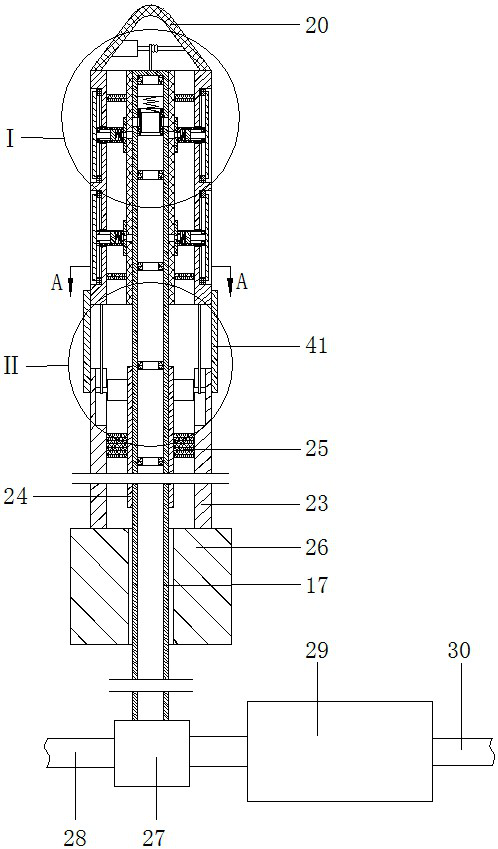
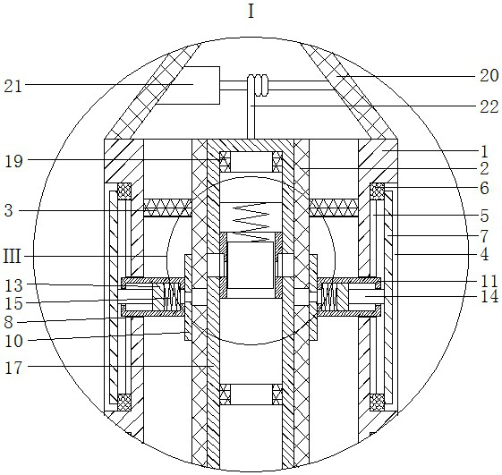
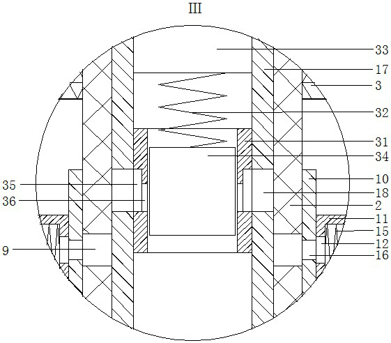
[0015]A surgical fixation device for visceral fixation in minimally invasive surgery, as shown in the figure, comprising a first outer tube 1 and a first inner tube 2, both of which are circular tubes, The first inner tube 2 is arranged in the first outer tube 1, the axes of the first inner tube 2 and the first outer tube 1 are collinear, and the ...


PUM


Abstract

A surgical fixation device for fixing organs in minimally invasive surgery comprises a first outer tube and a first inner tube. A first rectangular groove is arranged on the outer ring of the first outer tube. A sliding groove is arranged on the inner wall of the first rectangular groove. A sliding block is arranged in the sliding groove. An air bag is arranged on the outer wall of the first innertube. A cylinder is arranged on the air bag. A piston is arranged in the cylinder. A medical hose is arranged in the first inner tube. An electric covering mechanism is arranged on the medical hose.A guide cover is arranged on the first outer tube. A first servo motor is arranged in the guide cover. A second outer tube is arranged below the first outer tube. An angle adjusting mechanism is arranged between the first outer tube and the second outer tube. A handle is arranged at the lower end of the second outer tube. An electromagnetic change valve is arranged below the handle and provided with an air outlet tube. The electromagnetic change valve is installed at an air outlet end of an air pump. An air inlet tube is arranged at the air inlet end of the air pump. A power supply and a controller are arranged beside the air pump. According to the invention, a doctor can conveniently insert the surgical fixation device into the body of a patient, so that blocked organs can be stretched, stripped or fixed. The opening size stretched by a spring piece can be adjusted, so that organs of different sizes can be fixed.

Description

technical field [0001] The invention belongs to the field of minimally invasive surgical fixation devices, in particular to a surgical fixation device used for visceral fixation in minimally invasive operations. Background technique [0002] In minimally invasive surgery, surgeons often use large instruments to operate through small holes, and use endoscopic cameras to observe internal anatomical structures. Organs often interfere with the doctor's operation, so nurses are often required to use special surgical instruments to prop up, peel off, or fix these organs that have nothing to do with the lesion, so as to increase the operating space and facilitate the operation; However, the existing surgical instruments used to stretch or fix organs are too large to be easily inserted into minimally invasive incisions. Excessively large surgical instruments will also lead to excessively large surgical wounds, which is not conducive to postoperative recovery and aesthetics. Conten...

Claims


Application Information

Patent Timeline
Patent Type & Authority Applications(China)
IPC IPC(8): A61B17/02A61B90/17
CPCA61B17/02A61B90/17A61B17/0218A61B2017/0212
Inventor 万文兵
Owner THE SECOND AFFILIATED HOSPITAL TO NANCHANG UNIV
Learn More