
Blood vessel segmentation method and device and electronic equipment

A blood vessel and segmentation technology, applied in the field of medical images, can solve problems such as accurate segmentation of difficult blood vessels

Active Publication Date: 2021-02-02
赛诺威盛医疗科技(扬州)有限公司
View PDF13 Cites 5 Cited by
  • Summary
  • Abstract
  • Description
  • Claims
  • Application Information

AI Technical Summary

Problems solved by technology

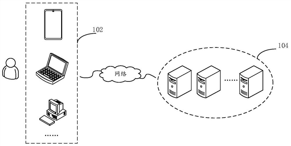
[0009] This application provides a blood vessel segmentation method, device and electronic equipment to solve the technical problem in the related art that it is difficult to accurately segment blood vessels with complex structures

Method used


Image

  • Blood vessel segmentation method and device and electronic equipment
  • Blood vessel segmentation method and device and electronic equipment
  • Blood vessel segmentation method and device and electronic equipment

Examples


Embodiment Construction

[0030]In order to enable those skilled in the art to better understand the solution of the present application, the technical solution in the embodiment of the application will be clearly and completely described below in conjunction with the accompanying drawings in the embodiment of the application. Obviously, the described embodiment is only It is an embodiment of a part of the application, but not all of the embodiments. Based on the embodiments in this application, all other embodiments obtained by persons of ordinary skill in the art without creative efforts shall fall within the scope of protection of this application.

[0031] It should be noted that the terms "first" and "second" in the description and claims of the present application and the above drawings are used to distinguish similar objects, but not necessarily used to describe a specific sequence or sequence. It is to be understood that the data so used are interchangeable under appropriate circumstances such ...


PUM


Abstract

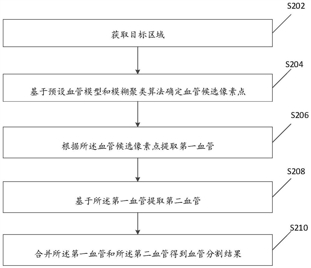
The invention provides a blood vessel segmentation method and device and electronic equipment, and the method comprises the steps of obtaining a target region which comprises a to-be-segmented region;determining blood vessel candidate pixel points based on a preset blood vessel model and a fuzzy clustering algorithm; extracting a first blood vessel according to the blood vessel candidate pixel points; extracting second blood vessels based on the first blood vessels, and the density of the first blood vessels is larger than that of the second blood vessels; and combining the first blood vesseland the second blood vessel to obtain a blood vessel segmentation result. According to the invention, the influence of other tissues with similar blood vessel characteristics on blood vessel segmentation can be effectively reduced, and the influence of blood vessel characteristics on blood vessel segmentation can be effectively reduced. The invention can improve precision of blood vessel segmentation.

Description

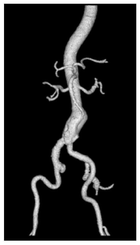
technical field [0001] The present application relates to the technical field of medical images, in particular to a blood vessel segmentation method, device and electronic equipment. Background technique [0002] Due to the complex structure of human blood vessels and the aging of the global population, there are many types of vascular diseases, and the incidence of vascular diseases is also increasing year by year. As a simple, rapid, non-invasive and safe method, multi-slice spiral CT angiography (CTA) is widely used in the diagnosis of vascular diseases. Separating and extracting the vascular structure from the CTA chest and abdomen data can assist doctors in observing the vascular structure, diagnosing vascular diseases and setting surgical plans. [0003] For vessels with complex structures, due to their complex structure, it brings great challenges to accurate segmentation, such as thoracoabdominal vessels, which have very complex structures, many vessels, and the sca...

Claims


Application Information

Patent Timeline
IPC IPC(8): G06T7/00G06T7/11G06T7/187G06T5/30G06K9/62
CPCG06T7/0012G06T7/11G06T7/187G06T5/30G06T2207/20032G06T2207/30101G06F18/23G06F18/22
Inventor 马双孙奇黄艳袁玉亮吴岩君
Owner 赛诺威盛医疗科技(扬州)有限公司
Learn More