Full-field OCT method and system for generating imaging of ocular fundus

A full-field, imaging technology, applied in the field of full-field OCT systems, can solve problems such as defocus and unclear images, and achieve the effect of eliminating smear

Active Publication Date: 2020-09-25
维索特克有限公司
View PDF12 Cites 0 Cited by
  • Summary
  • Abstract
  • Description
  • Claims
  • Application Information

AI Technical Summary

Problems solved by technology

Since the imaging properties of the refractive element are not the same in all eyes, without adjustment, a defocused and thus unclear image is produced on the image sensor

Method used


Image

  • Full-field OCT method and system for generating imaging of ocular fundus
  • Full-field OCT method and system for generating imaging of ocular fundus
  • Full-field OCT method and system for generating imaging of ocular fundus

Examples


Embodiment Construction

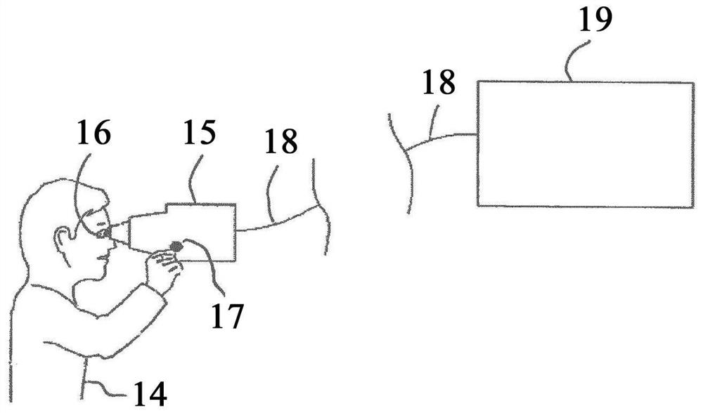
[0046] exist figure 1 In the full-field OCT system shown in accordance with the present invention, a patient 14 holds a handheld device 15 in his hand, producing images from the patient's fundus. The handheld device 15 is positioned in front of the patient 14's eyes 16 so that the patient 14 can see the fixation light inside the handheld device 15 . After the patient 14 sets its viewing direction according to the fixation light, the patient operates the switch 17, and the switch is used to trigger the imaging of the fundus.

[0047] After the recording, the image data are transmitted via the data network 18 to a central computer 19 remote from the patient 14 . The central computer 19 can be arranged, for example, in the center of a service provider running the field-wide OCT system. The system can be arranged such that the central computer 19 receives image data from a plurality of handheld devices 15 operating at different locations or in different installations. In partic...


PUM


Abstract

The invention relates to a full-field OCT method for generating an imaging of an ocular fundus (31), in which short-coherent light (22) is emitted and split into an object beam path (25) and a reference beam path (24). The object beam path (25) is directed onto the ocular fundus (33). The reference beam path (24) and a portion of the object beam path (25) reflected by the ocular fundus (31) are directed onto an image sensor (32), such that an interference between the reference beam path (24) and the object beam path (25) occurs on the image sensor (32), wherein the reference beam path (24) impinges on the image sensor (32) at an angle deviating from the object beam path (25). Before impinging on the image sensor (32), the reference beam path (24) impinges on an optical correction element (27) in order to reduce a chromatic aberration within the reference beam path (24). Intensity information and phase information is determined from a capturing of the image sensor. A focus-adjusted image of the ocular fundus is calculated. The invention also relates to a system that is suitable for carrying out the method. Images of the ocular fundus can be captured without the beam path being previously adapted to the refractive power of the eye lens.

Description

technical field [0001] The present invention relates to a full-field OCT method and a full-field OCT system for producing images of the fundus. Background technique [0002] The fundus of the human eye can be examined by optical coherence tomography (OCT). Capable of obtaining image information about tissue structures lying below the surface. In OCT measurements, short coherent light is split into an object beam path and a reference beam path. The object beam path is directed onto the fundus. A part of the object beam path reflected from the fundus interferes with the reference beam path. Information on the depth position of the scattering center in the fundus tissue can be obtained from the reference pattern. [0003] In classical OCT imaging, an object is probed (scanned) with an OCT beam to obtain a planar image (frontal image). Full-field OCT differs from these classical OCT methods in that an image sensor is used which takes a frontal image with one shot. [0004]...

Claims


Application Information

Patent Timeline
Patent Type & Authority Applications(China)
IPC IPC(8): A61B3/10
CPCA61B3/102G01B9/02091G01B9/02058G01B9/02032G01B9/02075G01B9/02083A61B3/1225
Inventor 彼得·科克格伦·许特曼赫尔格·祖德坎普亨德里克·斯帕尔迪克·希尔曼迈克尔·明斯特
Owner 维索特克有限公司
Why Eureka
Try Eureka