A non-invasive positioning device for ureteroscopic parapelvic cysts

A technology of flexible ureteroscope and positioning device, which is applied in application, medical science, diagnosis, etc., and can solve problems such as inconvenient application, inconvenient operation and use, and inconvenient operation and detection

Active Publication Date: 2022-07-26
HANGZHOU THIRD HOSPITAL
View PDF5 Cites 0 Cited by
  • Summary
  • Abstract
  • Description
  • Claims
  • Application Information

AI Technical Summary

Problems solved by technology

[0002] Parapelvic cyst is a relatively rare type of renal cystic disease. It is a non-hereditary disease. It mainly refers to cysts that occur near the renal pelvis. A certain calices is connected, so it is usually not easy to distinguish from renal cysts. Ultrasound three-push imaging can detect the integrity of the kidney from a 3D perspective because of its relatively primitive 2D imaging, which can provide better detection and judgment for the detection of renal pelvic cysts. However, ultrasound For 3D imaging, it is necessary to adjust the ultrasonic probe to the detection area with six degrees of freedom for detection and adjustment. The general method of handling is to support and clamp the probe through a mechanical arm structure capable of changing six degrees of freedom. However, in the During detection, the probe needs to be controlled to reciprocate in six degrees of freedom, which makes it extremely inconvenient for personnel to operate and detect
[0003] The notification number CN104095651A discloses a three-dimensional ultrasonic imaging system, including a vertical support, a gravity balance board for measuring the pressure distribution data information of the patient's feet, a computer, a second display, a support structure for supporting the patient, and a first display , ultrasonic scanner, and space sensor. The bracket has six degrees of freedom adjustment changes when the probe moves, and it is inconvenient for personnel to operate and use, so it cannot be conveniently applied in ultrasonic kidney detection

Method used


Image

  • A non-invasive positioning device for ureteroscopic parapelvic cysts
  • A non-invasive positioning device for ureteroscopic parapelvic cysts
  • A non-invasive positioning device for ureteroscopic parapelvic cysts

Examples


Embodiment Construction

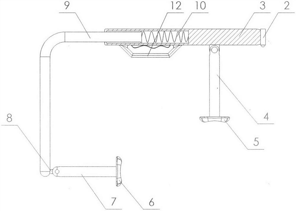
[0015] The present invention is a non-invasive locating device for ureteroscopic parapelvic cyst which is realized as follows: the present invention is a non-invasive locating device for ureteroscopic parapelvic cyst which consists of a data cable (1), a marking spotlight (2), a grip Holding rod (3), No. 1 connecting rod (4), No. 1 detector (5), No. 2 detector (6), No. 2 connecting rod (7), connecting block (8), bending rod (9) , a spring (10), an ultrasonic probe (11) and a holding handle (12), the holding rod (3) is divided into two sections from the middle, and one section is a hollow structure, the other end is a solid structure, the bending rod One end of (9) is sheathed in the hollow section of the holding rod (3), one end of the bending rod (9) is sheathed with a limiting ring, and the spring (10) is sheathed in the middle section of the holding rod (3). , one end of the spring (10) is connected with the holding rod (3), the other end of the spring (10) is connected wit...


PUM


Abstract

The present invention is a non-invasive positioning device for ureteroscope for renal pelvis cysts, which discloses a method for quickly positioning the No. 1 detection head above the patient's kidney by means of a marking spotlight, and using the No. 2 detector to detect the waist of the patient to form a three-dimensional image The detection device is characterized in that the holding rod is divided into two sections from the middle, and one section is a hollow structure, the other end is a solid structure, and one end of the bending rod is sleeved in the hollow section of the holding rod, and the folding One end of the bending rod is placed with a limiting ring, the spring is sleeved in the middle section of the holding rod, one end of the spring is connected with the holding rod, and the other end of the spring is connected with one end of the bending rod, marking the spotlight It is placed on the other end of the holding rod, with the shooting head facing down, a connecting block is fixed at the bottom of the solid section of the holding rod, and one end of the No. 1 connecting rod is clamped on the connecting block at the bottom of the holding rod, and passes The hinge shaft and the connecting block are hinged, and the No. 1 detector is placed on the other end of the No. 1 connecting rod.

Description

technical field [0001] The invention relates to a non-invasive positioning device for a renal pelvis cyst with a flexible ureteroscope, which relates to a device for rapid positioning and detection of a renal pelvis cyst, and belongs to the field of urology equipment. In particular, it relates to a detection device for quickly positioning the No. 1 detection head above the patient's kidney by identifying the spotlight, and performing detection on the patient's waist by the No. 2 detector to form a three-push image. Background technique [0002] Parapelvic cyst is a relatively rare type of renal cystic disease. It is a non-hereditary disease. It mainly refers to the cyst that occurs adjacent to the renal pelvis. In B-ultrasound detection, because the cyst is a round liquid dark area with A certain renal calyx is connected, and it is usually difficult to distinguish it from a renal cyst. However, because of its relatively primitive 2D imaging, three-push ultrasound imaging can...

Claims


Application Information

Patent Timeline
Patent Type & Authority Patents(China)
IPC IPC(8): A61B8/08A61B8/00
CPCA61B8/085A61B8/4254A61B8/4477
Inventor 诸靖宇李瑞鹏周鹏王彦彬
Owner HANGZHOU THIRD HOSPITAL
Why Eureka
Try Eureka