


Quick Research
Generate reliable direction feasibility study reports for your R&D in just a few steps.

Technical Q&A
Discover and master advanced knowledge NOW. Basics, ideas, possibilities, all at once.

Find Solutions
As an expert in R&D theories, this can generate solutions to your technical problems instantly.

Evaluate Feasibility
Analyze your overall solution with one click, know your potential R&D risks in advance.

Monitor Landscape
Get weekly tech updates, stay abreast of the latest tech innovations and key insights.
System and method for medical imaging
A technology of medical images, objects, applied in the field of medical imaging of the heart, analysis of medical images of the heart
- Summary
- Abstract
- Description
- Claims
- Application Information
AI Technical Summary
Problems solved by technology
Method used
Image


Examples
Embodiment 1
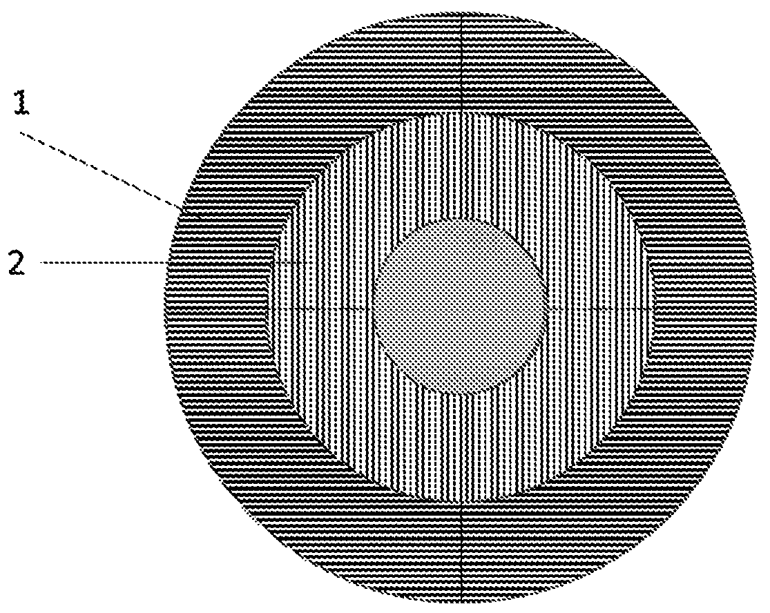
[0076] Detection of ischemia using the system of the invention
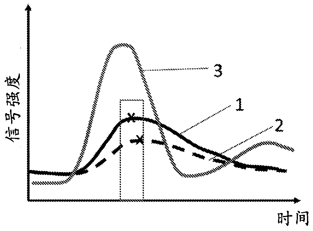
[0077] Figure 1A The epicardium of the heart is schematically illustrated, divided into an epicardial layer (1 ) and an endocardial layer (2) surrounding the central blood vessel. First pass CT images, MR images or SPECT images are taken in this direction and signals from individual layers (1, 2) are determined using the system of the invention. This generates many peaks such as Figure 1B with Figure 1C exemplified in . In these graphs, signal intensity-time at a reference point in the left ventricle is shown as a light gray peak (3). The signal intensity-time peak in the epicardial layer (1 ) is shown as a black solid line, and the signal intensity-time peak in the endocardial layer (2) is shown as a dashed line. Although the peak signal intensity in each of these layers can be expected to vary with time compared to the reference point, it can be expected that in a normal heart, the time to peak intens...
Embodiment 2
[0080] Determining the presence of scar tissue and distinguishing CAD from MVD
[0081] According to another aspect of the invention, the system is arranged to be based on measurements taken from a plurality of radial myocardial segments (4, 5, 6 and 7) in a plane similar to that of the slices used in Example 1. measure to get another metric such as Figure 2A exemplified in . In this case, the TTPI index, defined as the difference between the time to peak intensity at the reference point shown by the gray line (8) and the time to peak intensity in all individual radial segments, is at Figure 2B is relatively low, where the TTPI of the individual radial segments 4, 5, 6 and 7 are clearly similar. Where this result occurs after a positive ischemia identification as a result of the analysis of Example 1, this provides an indication that the individual may be suffering from MVD. Alternatively, where the result from Example 1 is negative, this result will confirm that the hea...
PUM
 Abstract
Description
Claims
Application Information
 - R&D Engineer
- R&D Manager
- IP Professional
- Industry Leading Data Capabilities
- Powerful AI technology
- Patent DNA Extraction
Browse by: Latest US Patents, China's latest patents, Technical Efficacy Thesaurus, Application Domain, Technology Topic, Popular Technical Reports.
© 2024 PatSnap. All rights reserved.Legal|Privacy policy|Modern Slavery Act Transparency Statement|Sitemap|About US| Contact US: help@patsnap.com