
Left ventricular myocardial segmentation method, device, and computer-readable storage medium

A left-ventricular, computer-programmed technology, used in the biomedical field, that solves problems that are time-consuming and require high levels of expert knowledge and experience

Inactive Publication Date: 2019-01-29
SHENZHEN INST OF ADVANCED TECH
View PDF7 Cites 7 Cited by
  • Summary
  • Abstract
  • Description
  • Claims
  • Application Information

AI Technical Summary

Problems solved by technology

Manual segmentation requires expert knowledge and experience and is time consuming

Method used


Image

  • Left ventricular myocardial segmentation method, device, and computer-readable storage medium
  • Left ventricular myocardial segmentation method, device, and computer-readable storage medium
  • Left ventricular myocardial segmentation method, device, and computer-readable storage medium

Examples


Embodiment Construction

[0035] In order to make the purpose, features and advantages of the present application more obvious and understandable, the technical solutions in the embodiments of the present application will be clearly and completely described below in conjunction with the drawings in the embodiments of the present application. Obviously, the described The embodiments are only some of the embodiments of the present application, but not all of them. Based on the embodiments in this application, all other embodiments obtained by those skilled in the art without making creative efforts belong to the scope of protection of this application.

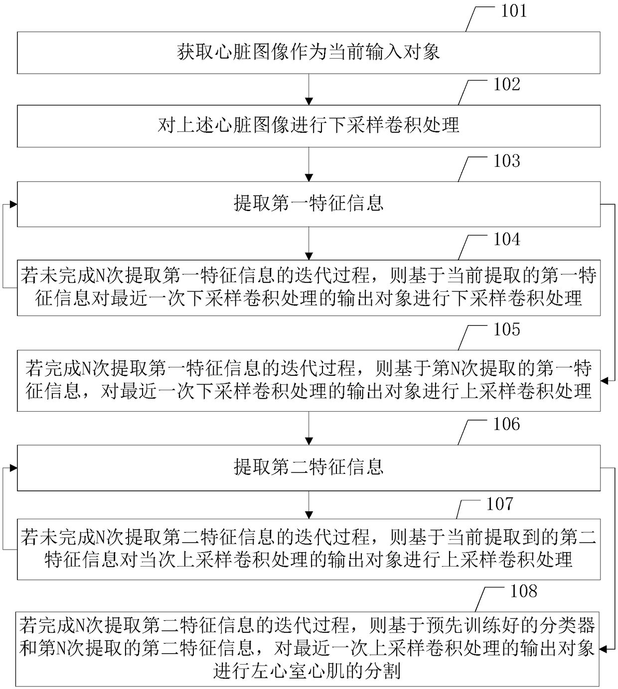
[0036] Such as Figure 1-a As shown, a method for segmenting left ventricular myocardium in the embodiment of the present application includes:

[0037] Step 101, acquiring heart images;

[0038] In an application scenario, step 101 may be expressed as: acquiring a cardiac image by using a magnetic resonance imaging (Magnetic Resonance Imaging, MRI) tec...


PUM


Abstract

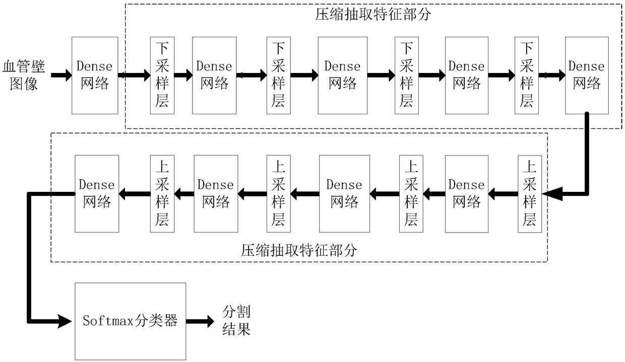
A left ventricular myocardial segmentation method, device and compute readable storage medium are disclosed, wherein, that left ventricular myocardial segmentation method comprises the follow steps of: obtaining a heart image; for the heart image, N times of down-sampling convolution processing and extraction of the first feature information are performed iteratively; if the iterative process of extracting the first feature information is completed for N times, performing N times of up-sampling convolution processing and extracting the second feature information for the output object of the Nth down-sampling convolution processing based on the first feature information extracted for the Nth time; if the iterative process of extracting the second feature N times is completed, the left ventricular myocardium is automatically segmented based on the pre-trained classifier. The technical proposal provided by the present application can effectively improve the efficiency of left ventricularmyocardial segmentation.

Description

technical field [0001] The present application relates to the field of biomedicine, and in particular to a left ventricular myocardium segmentation method, device and computer-readable storage medium. Background technique [0002] Segmentation of left ventricular myocardium is the main step in cardiac function analysis. Cardiac function analysis parameters such as left ventricular myocardial wall thickness and ejection volume are all based on accurate segmentation of left ventricular myocardium. The gray scale of the left ventricular myocardium is similar to that of the background, and the papillary muscle and trabeculae in the left ventricle belong to the same substance as the myocardium, and there are artifacts. Therefore, left ventricular myocardium segmentation is a difficult but important task. [0003] Currently, methods for left ventricular myocardium segmentation mainly use magnetic resonance imaging (Magnetic Resonance Imaging, MRI) technology to obtain heart image...

Claims


Application Information

Patent Timeline
IPC IPC(8): G06T7/10G06K9/46G06K9/62G06N3/04
CPCG06T7/10G06T2207/30048G06T2207/10088G06T2207/10132G06T2207/20081G06V10/462G06N3/045G06F18/241
Inventor 郑海荣刘新胡战利李思玥吴垠梁栋杨永峰
Owner SHENZHEN INST OF ADVANCED TECH
Learn More