
Training device for endoscopic endonasal skull base surgery

A technology of training equipment and endoscope, which is applied in the field of training equipment, can solve the problems of not finding training equipment and training difficulties

Inactive Publication Date: 2017-12-01
ONO
View PDF9 Cites 1 Cited by
  • Summary
  • Abstract
  • Description
  • Claims
  • Application Information

AI Technical Summary

Problems solved by technology

This presents difficulties in training to deal with injuries to the internal carotid artery
No literature was found on the device used for training in endoscopic transnasal skull base surgery

Method used


Image

  • Training device for endoscopic endonasal skull base surgery
  • Training device for endoscopic endonasal skull base surgery
  • Training device for endoscopic endonasal skull base surgery

Examples


Embodiment Construction

[0033] A preferred embodiment of a training device for transnasal endoscopic skull base surgery configured according to the present invention will now be described in detail with reference to the accompanying drawings.

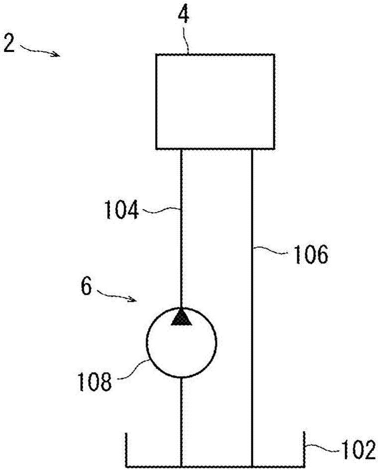
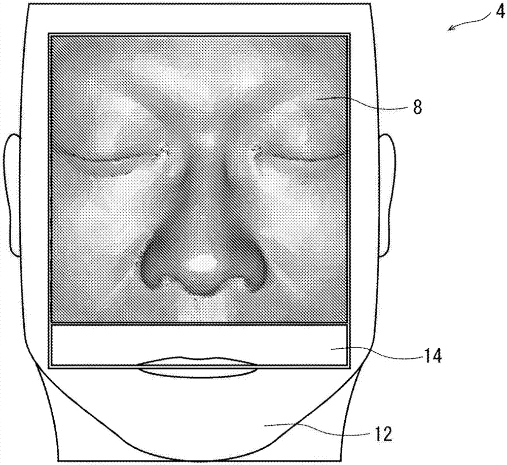
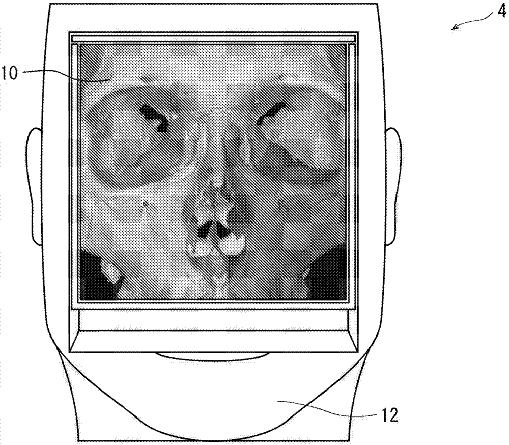
[0034] Such as figure 1 As shown, the training equipment for transnasal endoscopic skull base surgery is represented by numeral 2 as a whole and consists of a human head model 4 and a fluid circulation component 6 . In the illustrated embodiment, the human head model 4 includes a front portion 8, a rear portion 10, a base block 12 and wedges 14, as figure 2 and 3 shown.

[0035] Figure 4 and 5 will be referenced for illustration. A front portion 8 that reproduces at least a part of a human face is provided on the front surface of the human head model 4, and importantly, includes a nose portion 16 formed with nostrils 16a. In the illustrated embodiment, the front part 8 formed in a quadrangular shape when viewed from the front, which is separated from t...


PUM


Abstract

Provided is a device for carrying out training for endoscopic endonasal skull base surgery. A training device 2 for endoscopic endonasal skull base surgery is constituted of a human head model 4 and a liquid circulation means 6. The human head model 4 is provided with a front part 8 that includes a nose part 16 in which nostrils 16a are formed and a main part 40 that includes at least part of a nasal septum 44, at least part of a nasal cavity lateral wall 42, at least part of a sphenoidal sinus posterior wall 66, and at least part of an internal carotid artery 72. The liquid circulation means 6 is provided with: a retention tank 102 for retaining a nontransparent colored liquid; a supply flow path 104 disposed between the retention tank 102 and the at least part of an internal carotid artery 72; a return flow path 106 disposed between the at least part of an internal carotid artery 72 and the retention tank 102; and a circulating pump 108 for circulating the liquid in the retention tank 102 through the supply flow path 104, the at least part of an internal carotid artery 72, and the return flow path 106.

Description

technical field [0001] The present invention relates to a training device for transnasal endoscopic surgery of skull base lesions. Background technique [0002] In the sphenoid bone located near the center of the human skull base, a depression called sella exists near its center. The pituitary gland, the hormone-secreting endocrine organ, is housed in this depression. Tumors arising in the pituitary gland have so far undergone transsphenoidal sinus surgery, which involves cutting the upper gingival part (upper tooth root) behind the upper lip, and approaching the tumor from the incision under a microscope. However, according to improvements in miniaturized surgical instruments including endoscopes, transnasal endoscopic surgery has begun to be performed on pituitary tumors, and compared with transsphenoidal sinus surgery performed under a microscope, transnasal endoscopic surgery provides The wide field of view allows the site of surgery to be clearly identified and is als...

Claims


Application Information

Patent Timeline
Patent Type & Authority Applications(China)
IPC IPC(8): G09B9/00B33Y80/00G09B23/28
CPCB33Y80/00G09B23/285G09B9/00B33Y10/00B33Y50/02B29C64/153B29L2031/7546G09B23/303G09B23/34
Inventor 大野秀则武藤淳
Owner ONO
Learn More