
Split Proctoscope Ultrasound Doppler Probe

A proctoscope and split-type technology, applied in the field of medical devices, can solve the problems of single function, suture and suspension operation of hemorrhoids, etc., and achieve the effects of simple connection, heavy weight and clear vision.

Active Publication Date: 2016-06-22
南京奥珂森电子有限公司
View PDF6 Cites 0 Cited by
  • Summary
  • Abstract
  • Description
  • Claims
  • Application Information

AI Technical Summary

Problems solved by technology

Therefore, the function of the original probe showed certain limitations. Because the window was too small, it was impossible to suture and suspend the hemorrhoid body, and there was a problem of single function, which could not meet the requirements of modern treatment technology.

Method used


Image

  • Split Proctoscope Ultrasound Doppler Probe
  • Split Proctoscope Ultrasound Doppler Probe
  • Split Proctoscope Ultrasound Doppler Probe

Examples


Embodiment 1

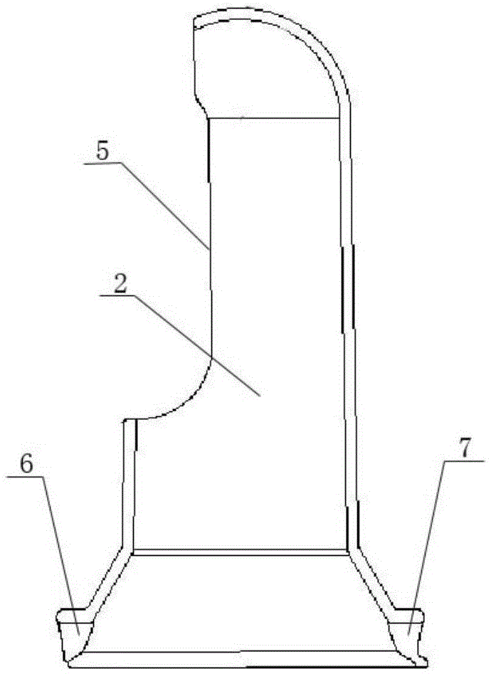
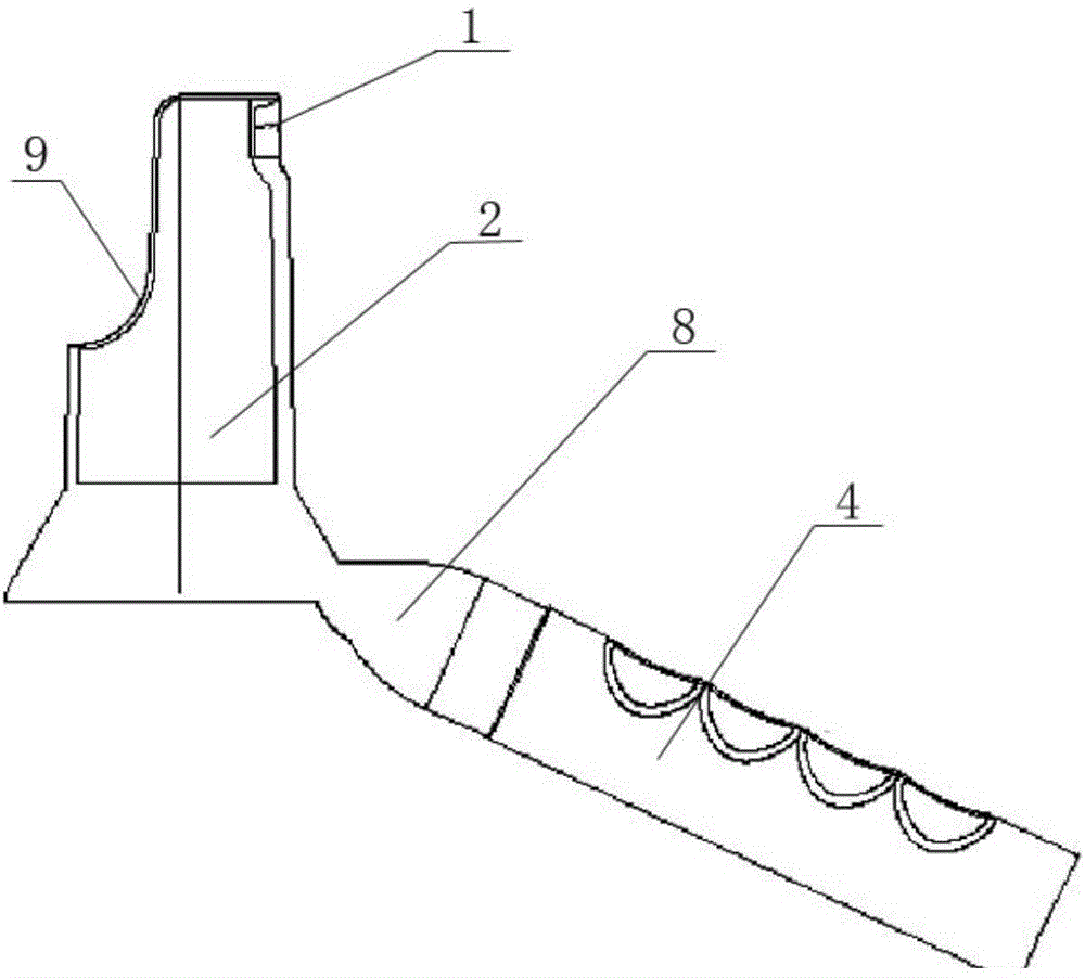
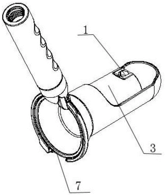
[0018] This embodiment provides a split type rectoscope ultrasound Doppler probe, such as Figure 1 to Figure 4 As shown, it includes an ultrasonic transducer 1 installed on the casing, the casing includes an outer casing 2, an inner casing 3 and a handle 4 connected to the inner casing, the handle 4 is equipped with an ultrasonic transducer connector socket, and the outer casing is one end Closed tubular body, the closed end of the jacket is a hemispherical structure, the side of the tubular body is provided with a ligation window 5, the length of the ligation window 5 is 1 / 2 of the length of the jacket, and the width of the ligation window is 1 / 3 of the width of the jacket; The unclosed end is the operation operation port, and the edge of the operation operation port is provided with ligation positioning slots 6 and suture positioning slots 7 which are symmetrically distributed, and the ligation positioning slots 6 and the ligation window 5 are on the same side of the tubular...


PUM


Abstract

The invention relates to a split-type rectoscope ultrasonic Doppler probe, which includes an ultrasonic transducer installed on a casing, the casing includes an outer casing, an inner casing and a handle connected to the inner casing, and the outer casing is a tubular body with one end closed. The side of the tubular body is provided with a ligation window, the unclosed end of the tubular body is the operation port, and the edge of the operation port is provided with a ligation positioning slot and a suture positioning slot symmetrically distributed, the ligation positioning slot and the ligation window are in the The same side of the tubular body; the inner sleeve is two ends through the tube body, the length of the inner sleeve is less than the length of the outer sleeve, one end of the inner sleeve body is equipped with an ultrasonic transducer, and the other end is provided with an interface for installing a handle. The transducer is arranged on the same side of the inner casing body, and a suturing window is arranged on the inner casing body on the opposite side where the ultrasonic transducer is installed. The probe of the present invention adopts a split structure, which not only retains the ligation function of the original Doppler probe, but also designs a split structure of the probe through creative design, which can form two operating stations, thereby realizing ligation and suspension of hemorrhoids Suture ligation can be performed under the same probe.

Description

technical field [0001] The invention relates to an ultrasonic Doppler probe, in particular to a split-type rectoscope ultrasonic Doppler probe, which belongs to the technical field of medical instruments. Background technique [0002] At present, when the existing rectoscope ultrasonic Doppler probe is used to treat hemorrhoids, it can detect the position of hemorrhoid arteries, and then ligate the hemorrhoid arteries to treat hemorrhoids. [0003] With the advancement of medical technology and the improvement of surgical requirements, in the latest hemorrhoid treatment technology, not only the hemorrhoid arteries should be inspected, the hemorrhoid arteries should be ligated, but also the hemorrhoids should be sutured and suspended. Therefore, the function of the original probe has shown certain limitations, because the window is too small to suture and suspend the hemorrhoid, and there is a problem of single function, which cannot meet the requirements of modern treatment ...

Claims


Application Information

Patent Timeline
Patent Type & Authority Patents(China)
IPC IPC(8): A61B17/12A61B17/04A61B8/12
CPCA61B8/12A61B17/0469A61B17/12013
Inventor 钱旭波
Owner 南京奥珂森电子有限公司
Learn More