
Protective film for ultrasonic probe

A technology of ultrasonic probe and protective film, which is applied in ultrasonic/sonic/infrasonic diagnosis, acoustic diagnosis, infrasonic diagnosis, etc. It can solve problems such as shortened service life and patient pain, and achieve the effects of prolonging service life, relieving pain and saving time

Inactive Publication Date: 2013-01-30
滕学东
View PDF9 Cites 1 Cited by
  • Summary
  • Abstract
  • Description
  • Claims
  • Application Information

AI Technical Summary

Problems solved by technology

[0003] The invention aims at the disadvantages of the prior art that the ultrasonic probe brings great pain to the patient when it is used, and that frequent disinfection will shorten its service life, and provides a protective film for the ultrasonic probe

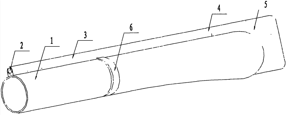
Method used


Image

  • Protective film for ultrasonic probe

Examples


Embodiment Construction

[0013] The principles and features of the present invention are described below in conjunction with the accompanying drawings, and the examples given are only used to explain the present invention, and are not intended to limit the scope of the present invention.

[0014] A protective film for an ultrasonic probe, comprising a body 1, which is a film sleeve with one end closed; the outer wall of the body 1 is provided with a needle track 3 and a fastening belt 6, and an image colloid 5 is provided at the closed end; The needle track 3 includes a needle inlet port 2 and a needle outlet port 4, and the needle outlet port 4 is connected with the image colloid 5.

[0015] When in use, the present invention is directly placed on the ultrasonic probe, fastened on the ultrasonic probe with the fastening band 6, and the puncture biopsy needle is inserted into the needle channel 3 from the needle inlet 2, so that the needle is located at the needle outlet 4, Then place the ultrasonic p...


PUM


Abstract

The invention relates to protective films, in particular to a protective film for an ultrasonic probe, belongs to the technical field of aspiration biopsy of ultrasonic departments, and overcomes the defects that the ultrasonic probe brings a lot of pains for patients during use and service life of the ultrasonic probe is shortened due to constant sterilization in the prior art. The protective film for the ultrasonic probe comprises a body which is a film sleeve with a closed end. A needle passage and a fastening strap are arranged on the outer wall of the body. Image colloid is disposed at the closed end. The needle passage comprises a needle inlet and a needle outlet. The needle outlet is connected with the image colloid. The protective film for the ultrasonic probe is mainly mounted on the ultrasonic probe for aspiration biopsy of prostate glands. The protective film for the ultrasonic probe is also applicable to gynecological puncture operations for treating vaginal cyst.

Description

technical field [0001] The invention relates to a protective film, in particular to an ultrasonic probe protective film, which belongs to the technical field of ultrasonic puncture biopsy. Background technique [0002] At present, in the traditional prostate biopsy method, the first step is to launch the biopsy gun after seeing the needle tip on the real-time ultrasound image, that is to say, the needle tip must be pierced into the human body before it can be seen, and this process is performed in the rectum. The surgeon cannot see it with his eyes, so he needs to slowly insert the needle and observe the position of the needle tip on the ultrasound image. An important issue involved at this time is that when a relatively thick puncture biopsy needle is slowly advanced in the human body, the patient will feel obvious pain, even unbearable; the second step is to aim the needle tip at the puncture target, and use the biopsy gun to launch the prostate puncture biopsy At this ti...

Claims


Application Information

Patent Timeline
Patent Type & Authority Applications(China)
IPC IPC(8): A61B8/00
Inventor 滕学东
Owner 滕学东
Learn More