Instrument for sectioning and dyeing biological sample on electronic microscope

A technology of electron microscopy and biological samples, applied in the preparation of test samples, etc., can solve the problems of biological sample pollution, difficult precision, pollution, etc., and achieve the effects of eliminating pollutants, reducing pollutants, and reducing time lag

Inactive Publication Date: 2015-06-17
FUDAN UNIV
View PDF4 Cites 0 Cited by
  • Summary
  • Abstract
  • Description
  • Claims
  • Application Information

AI Technical Summary

Problems solved by technology

Conventional methods of dyeing biological samples after embedding and sectioning are usually purely manual, because the time difference during lead staining cannot be controlled, and the accuracy is difficult to grasp, and the precipitation caused by the reaction of lead staining solution with carbon dioxide in the air will pollute biological samples. As a result, high-quality experimental results cannot be obtained; and because the original staining method is relatively cumbersome, block dyeing is often used when staining biological samples (uranyl acetate), and at least 1ml of uranyl acetate dye solution is required to prepare a sample. Thereby also correspondingly caused the waste and the pollution of uranyl acetate solution

Method used


Image

  • Instrument for sectioning and dyeing biological sample on electronic microscope

Examples


Embodiment 1

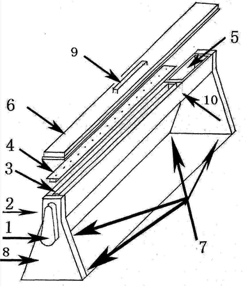
[0031] Such as figure 1As shown, the instrument in the present invention utilizes the positioning flap plus positioning lock function technology to perform multiple staining (uranyl acetate staining, lead staining, negative staining) before observing the electron microscope sample, and the described instrument is provided with a lock Control the dyeing instrument with a built-in flap and a groove that can isolate air circulation, which consists of a positioning handle 1, a water outlet 2, a groove flap 3, a slat 4, a built-in lidless box 5, a top cover 6, and a stabilizing gasket 7 and the groove frame 8; the groove flap 3 is placed in the top groove of the groove frame 8, and the positioning handle 1 is arranged on the outside of the groove frame 8, and its position corresponds to the groove flap 3. The slat 4 is placed above the groove flap 3, the built-in coverless box 5 is located in the top groove of the groove frame 8, one side of the groove flap 3, and the top cover 6 i...


PUM

PropertyMeasurementUnit
lengthaaaaaaaaaa
lengthaaaaaaaaaa
widthaaaaaaaaaa

Abstract

The invention relates to an instrument for sectioning and dyeing a biological sample on an electronic microscope, which belongs to the field of biological sample preparation. The instrument is composed of a locating handle, a water outlet, a groove flap board, a wattle, a built-in coverless box, a top cover, a stable gasket and a groove frame, wherein the groove flap board is arranged in a top groove of the groove frame; the locating handle is arranged on the outer side of the groove frame; the wattle is arranged above the groove flap board; the built-in coverless box is located in the top groove of the groove frame and arranged on one side of the groove flap board; the top cover is covered on the top groove of the groove frame; and the bottom part of the groove frame is provided with the stable gasket. According to the invention, the instrument can perform dyeing on biological sample parity for many times without repeated manual operations; thus, the time difference can be reduced; the generation of pollutant is eliminated; a high-quality experiment result is ensured to obtain; and the instrument can be used for immunohistochemical incubation of a histochemical microscope and preparation of macromolecule sample drop tablets and the like.

Description

technical field [0001] The invention belongs to the field of biological sample preparation, and in particular relates to an instrument for staining slices of biological samples under an electron microscope. Background technique [0002] Since the birth of the electron microscope, people have been continuously exploring and improving the pre-preparation, sectioning, and staining techniques of biological samples for electron microscope observation in order to obtain higher-quality real images and experimental results. Conventional methods of dyeing biological samples after embedding and sectioning are usually purely manual, because the time difference during lead staining cannot be controlled, and the accuracy is difficult to grasp, and the precipitation caused by the reaction of lead staining solution with carbon dioxide in the air will pollute biological samples. As a result, high-quality experimental results cannot be obtained; and because the original staining method is re...

Claims


Application Information

Patent Timeline
Patent Type & Authority Patents(China)
IPC IPC(8): G01N1/31
Inventor 高鸿建
Owner FUDAN UNIV
Why Eureka
Try Eureka