Neuroprotective Ganoderma compositions and methods of use
A technology of Ganoderma lucidum extract and Ganoderma lucidum, applied in] in one field, can solve the problems of weakening the inflammatory response of microglial cells, no report of Ganoderma lucidum, etc.
- Summary
- Abstract
- Description
- Claims
- Application Information
AI Technical Summary
Problems solved by technology
Method used
Image
Examples
preparation example Construction
[0063] MES 23.5 Preparation of Cell Membrane Fractions
[0064] After MES 23.5 cells were exposed to MPP+10 μM for 24 hours, they were harvested in a medium containing 0.25M sucrose, 100mM PBS, 1mM MgCl 2 , 1mM EDTA and 2μM protease inhibitor PMSF buffer, and use glass-Teflon homogenizer (glass-teflon homogenizer) to homogenize it (Le, W.D.et al., 2001, J.Neurosci., 21: 8447-8455). The homogenate was then centrifuged at 8000 xg for 10 minutes at 4°C to remove the native nuclear fraction. The supernatant was centrifuged again at 100000 x g for 60 min at 4 °C. The pellet was homogenized and suspended in culture medium and used as the neuronal membrane fraction.
[0065] High affinity [3H] dopamine uptake assay
[0066] With 1ml Krebs-Ringer buffer (16mM NaH 2 PO 4 , 16mM Na 2 HPO 4 , 119mM NaCl, 4.7mM KCl, 1.8mM CaCl 2 , 1.2 mM MgSO 4 , 1.3 mM EDTA and 5.6 mM glucose; pH 7.4) washed the cells in each well. The cells were then incubated with 10 nM [3H]dopamine in Krebs...
Embodiment 1
[0078] Example 1-LPS and MPP+ treated dopaminergic membrane-induced microglia activation
[0079]To model microglial activation in neurodegeneration, LPS and MPP were used in microglial cultures or co-cultures of dopaminergic neurons (MES23.5 cell line) and microglia + Treated dopaminergic cell membranes serve as stimuli.
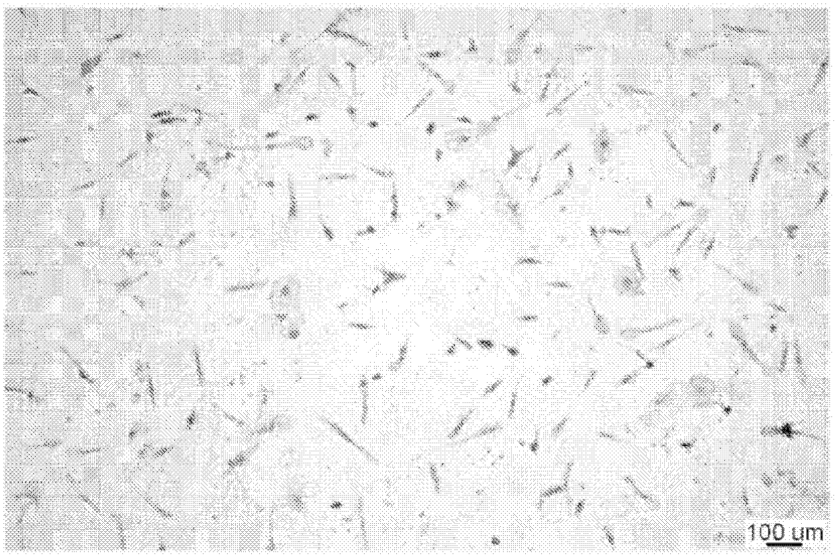
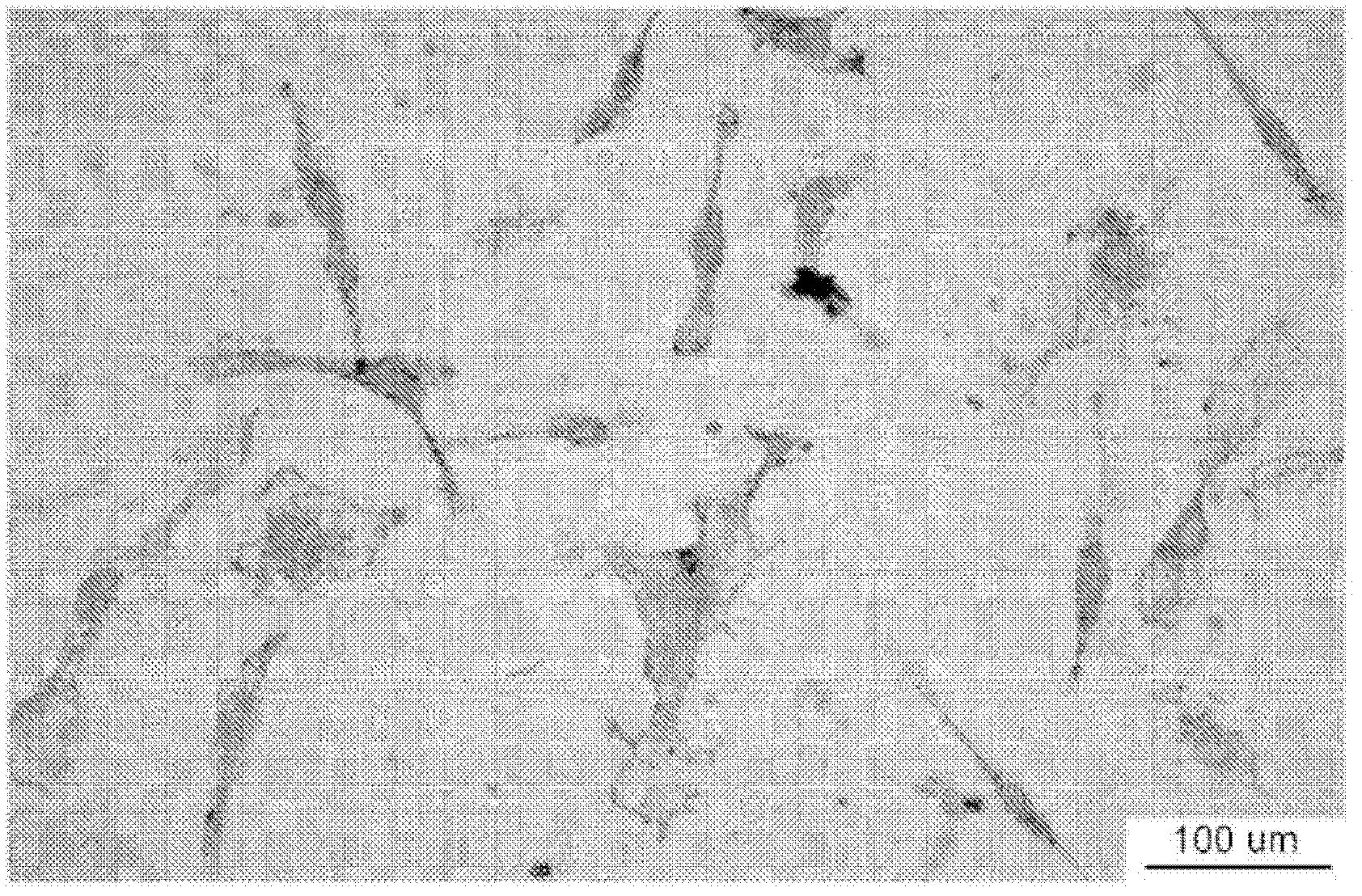
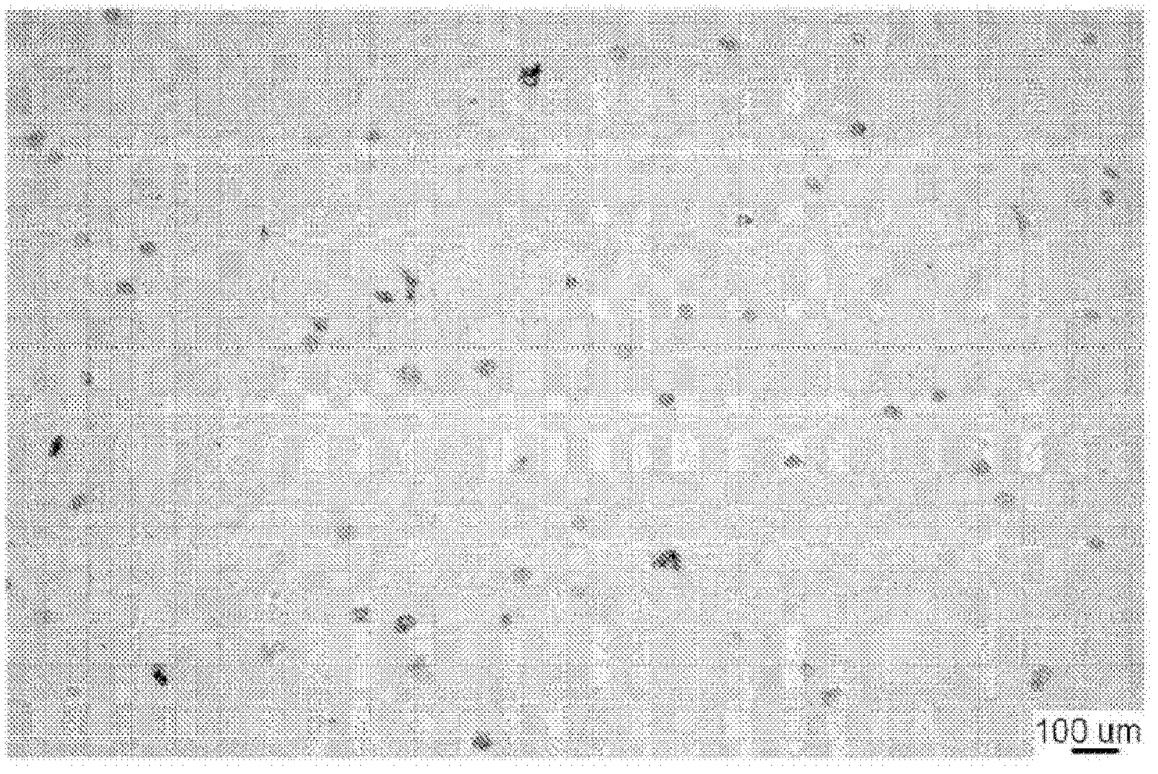
[0080] Microglia were visualized by staining for the CR3 complement receptor using the monoclonal antibody OX-42. The purity of the microglia culture was -95%. Non-activated microglia displayed branched shapes or bipolar or multipolar protrusions (Fig. 1a and b). Activated microglia exhibited an amoeboid-like morphology (Fig. 1c and d).
[0081] Among numerous neurotoxic factors, NO, TNF-α, IL-1β, and superoxide may be the main regulators of dopaminergic neurodegeneration induced by microglial activation. LPS-induced microglial activation was characterized by measuring TNF-α and IL-1β levels, two well-documented cytokines that reflect microglial activat...
Embodiment 2
[0085] Example 2 - Ganoderma lucidum prevents the production of pro-inflammatory factors and ROS derived from microglia
[0086] Microglia can produce cytokines as a result of activation (20-22). To elucidate the underlying mechanism of the neuroprotective activity of Ganoderma lucidum, the effects of Ganoderma lucidum on the levels of microglia-derived inflammatory cytokines and ROS were investigated. Microglial cultures were pretreated with different doses (50–400 μg / ml) of Ganoderma lucidum for 30 min before exposure to LPS or MPP + Processed CF.
[0087] Such as image 3 and 4 It was shown that low doses (50 μg / ml) of G. lucidum had minimal inhibitory effects, whereas pretreatment with higher doses (100–400 μg / ml) of G. lucidum potently reduced NO and CF induced by LPS or CF in a concentration-dependent manner. Increase of SOD.
[0088] At the same concentration, Ganoderma lucidum also significantly reduced exposure to LPS and MPP + Release of TNF-[alpha] and IL-1[be...
PUM
 Abstract
Description
Claims
Application Information
 - R&D
- Intellectual Property
- Life Sciences
- Materials
- Tech Scout
- Unparalleled Data Quality
- Higher Quality Content
- 60% Fewer Hallucinations
Browse by: Latest US Patents, China's latest patents, Technical Efficacy Thesaurus, Application Domain, Technology Topic, Popular Technical Reports.
© 2025 PatSnap. All rights reserved.Legal|Privacy policy|Modern Slavery Act Transparency Statement|Sitemap|About US| Contact US: help@patsnap.com


