
Fluorescence immunochromatographic assay and kit for quantitatively detecting N-terminal pro brain natriuretic peptide

A fluorescence immunochromatography and quantitative detection technology, applied in the field of medical testing, can solve the problems of low sensitivity, expensive instruments, and inaccurate quantification, and achieve the effect of improving detection sensitivity and sensitivity.

Active Publication Date: 2012-07-11
SHENZHEN KANGMEI BIOTECH
View PDF4 Cites 20 Cited by
  • Summary
  • Abstract
  • Description
  • Claims
  • Application Information

AI Technical Summary

Problems solved by technology

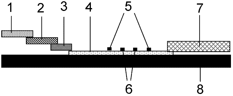
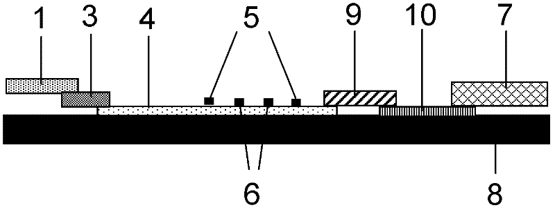
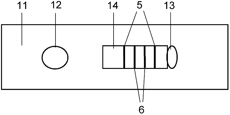
[0012] The technical problem to be solved in the present invention is to provide a fluorescent immunochromatographic method for quantitatively detecti

Method used


Image

  • Fluorescence immunochromatographic assay and kit for quantitatively detecting N-terminal pro brain natriuretic peptide
  • Fluorescence immunochromatographic assay and kit for quantitatively detecting N-terminal pro brain natriuretic peptide
  • Fluorescence immunochromatographic assay and kit for quantitatively detecting N-terminal pro brain natriuretic peptide

Examples


Embodiment 1

[0070] Example 1: Quantitative detection of NT-proBNP by modifying antibodies to quantum dots by chemical cross-linking and using direct pre-wetting immunochromatography

[0071] (1) Modification of quantum dots and antibodies

[0072] Add 60pmol quantum dots with an emission wavelength of 650nm, 10μg EDC, 15μg NHS solution and 10-30μg goat anti-human NT-proBNP monoclonal antibody solution to pH 7.4 phosphate buffer solution, mix well and react at room temperature for 4h, add 1mg glycine to block . Separating and purifying with a chromatographic column or a chromatographic column to obtain NT-proBNP antibody-modified quantum dots. Similarly, quantum dots modified with goat anti-rabbit antibody were obtained. The fluorescence emission wavelength of the quantum dot modified by the NT-proBNP antibody is 650nm, and the fluorescence emission wavelength of the quantum dot modified by the goat anti-rabbit antibody is 570nm.

[0073] (2) Construction of the kit

[0074] Mix the ab...

Embodiment 2

[0082] Example 2: Quantitative detection of NT-proBNP by modifying antibodies to quantum dots by biotin-avidin method and using indirect pre-wetting immunochromatography

[0083] (1) Synthesis and modification of quantum dots

[0084] According to the method of Example 1, streptavidin-modified quantum dots were prepared. Similarly, quantum dots modified with goat anti-rabbit antibody were obtained.

[0085] Dilute NT-proBNP monoclonal antibody to 1mg / ml with 0.1mol / L sodium bicarbonate buffer (pH 8.0), dissolve 1mg N-hydroxysuccinimide biotin (NHSB) with 1ml dimethyl sulfoxide (DMSO), Take 1ml monoclonal antibody solution and add 20-120μl NHSB solution, and react at room temperature for 4 hours. Add 1mol / L NH 4 Cl, stirred at room temperature for 10 min. Purify by ultrafiltration centrifugation to remove free biotin to obtain biotinylated mAb.

[0086] Streptavidin-modified quantum dots and biotinylated monoclonal antibodies are mixed at a ratio of 1:3 to 1:12 to obtain N...


PUM

PropertyMeasurementUnit
Emission wavelengthaaaaaaaaaa

Abstract

The invention discloses a fluorescence immunochromatographic assay and kit for quantitatively detecting acute myocardial infarction marker, namely, N-terminal pro brain natriuretic peptide (NT-proBNP). The fluorescence immunochromatographic assay for quantitatively detecting the NT-proBNP realizes fluorescence quantitative detection on the basis of optimizing various constituent parts of test paper by using excellent fluorescence characteristic of quantum dots and combining a bicolor marking technology and an immunochromatographic technology. Compared with the conventional colloidal gold immunochromatographic assay, the fluorescence immunochromatographic assay disclosed by the invention has the advantages of good marking stability, low non-specificity, high sensitivity, wide linear range and quantifying accuracy. The fluorescence immunochromatographic kit disclosed by the invention is used for carrying out quantitative detection on the NT-proBNP and detecting whole blood, blood serum and blood plasma samples, can provide reference for diagnosis of cardiovascular and cerebrovascular diseases and can be widely applied to primary hospitals and clinics.

Description

technical field [0001] The invention relates to the field of medical testing, in particular to a fluorescent immunochromatographic method for quantitatively detecting nitrogen-terminal brain natriuretic peptide NT-proBNP and a kit thereof. Background technique [0002] In 1988, Japanese scholar Tetsuji Sudoh first isolated a polypeptide with strong natriuretic, diuretic, vasodilator and hypotensive effects from pig brain, named BNP1. Subsequent studies have shown that not only BNP is a polypeptide, but a group of polypeptides are gradually developed and produced in the process of biological evolution (including at least ANP, BNP, CNP and DNP in humans and vertebrates), called natriuretic peptides (NP) family. Its function is to maintain the homeostasis of volume, osmolarity, and pressure regulation of the circulatory system. A large number of basic and clinical studies have shown that the level of BNP in the blood increases significantly in heart failure. As a new biomarke...

Claims


Application Information

Patent Timeline
IPC IPC(8): G01N33/68G01N21/64
Inventor 王东
Owner SHENZHEN KANGMEI BIOTECH
Learn More