Low dose single step grating based X-ray phase contrast imaging

An X-ray and grating technology, applied in the field of X-ray phase contrast imaging, to achieve the effect of reducing the dose

Inactive Publication Date: 2013-07-10
INST OF HIGH ENERGY PHYSICS CHINESE ACAD OF SCI +1
View PDF6 Cites 9 Cited by
  • Summary
  • Abstract
  • Description
  • Claims
  • Application Information

AI Technical Summary

Problems solved by technology

[0007] Clearly, this method is burdened with limitations both in the (long) data acquisition time and in the stringent dose delivered to the specimen

Method used


Image

  • Low dose single step grating based X-ray phase contrast imaging
  • Low dose single step grating based X-ray phase contrast imaging
  • Low dose single step grating based X-ray phase contrast imaging

Examples


Embodiment Construction

[0046] With reference to the above drawings, an innovative method for extracting phase information is proposed, which does not require a stepping process, thus overcoming the limitations of data acquisition time and dose released to the specimen.

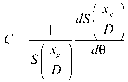
[0047] This novel method relies on the physical similarity between the crystal analyzer-based system and the grating interferometer. Both technologies record the refraction angle signal and are similar to the rocking curve of a crystal analyzer. The nature of the offset curve (see Figure 1) can be used to fully describe the performance of the grating interferometer. The refraction angle, the phase information of the sample, can be extracted by setting a grating interferometer in the center position where the intensity curve follows the linear behavior.

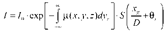
[0048] According to the previous simulation, the intensity recorded by the detector positioned behind the grating interferometer I It can be expressed as:

[0049]

[0050] Where μ is the...


PUM


Abstract

Phase sensitive X-ray imaging methods provide substantially increased contrast over conventional absorption based imaging, and therefore new and otherwise inaccessible information. The use of gratings as optical elements in hard X-ray phase imaging overcomes some of the problems impairing the wider use of phase contrast in X-ray radiography and tomography. To separate the phase information from other contributions detected with a grating interferometer, a phase-stepping approach has been considered, which implies the acquisition of multiple radiographic projections. Here, an innovative, highly sensitive X-ray tomographic phase contrast imaging approach is presented based on grating interferometry, which extracts the phase contrast signal without the need of phase stepping. Compared to the existing phase step approach, the main advantage of this new method dubbed “reverse projection” is the significantly reduced delivered dose, without degradation of the image quality.

Description

Technical field [0001] The invention relates to a method and system for X-ray phase contrast imaging based on a low-dose single-step grating. Background technique [0002] As we all know, unlike traditional visible light optics, the refractive index in X-ray optics is very close to and less than one, because the energy of X-ray photons is often much greater than the energy of atomic resonance. In the first approximation, for small and negligible anisotropy in the medium, the complex form of the refractive index can be used to express the refractive index that characterizes the optical properties of the tissue, including X-ray absorption: , Where δ is the reduction of the real part of the refractive index, which characterizes the phase shift properties, and the imaginary part β describes the absorption properties of the sample. In traditional absorption-based radiography, X-ray phase shift information is usually not directly used for image reconstruction. However, where the phot...

Claims


Application Information

Patent Timeline
Patent Type & Authority Patents(China)
IPC IPC(8): A61B6/00G01N23/04
CPCA61B6/484A61B6/4291G01N23/04A61B6/00G01N23/041
Inventor 朱佩平吴自玉M.斯塔姆帕诺尼
Owner INST OF HIGH ENERGY PHYSICS CHINESE ACAD OF SCI
Why Eureka
Try Eureka