Processing method for space-occupying lesion ultrasonic images

A technology of ultrasonic image and processing method, which is applied in the directions of ultrasonic/sonic/infrasonic image/data processing, image data processing, image analysis, etc., which can solve the problems of poor reproducibility of results, easy misdiagnosis, missed diagnosis, time-consuming and labor-intensive problems, etc. Achieve improved objectivity and better diagnosis

Inactive Publication Date: 2012-10-03
SHENZHEN UNIV
View PDF0 Cites 0 Cited by
  • Summary
  • Abstract
  • Description
  • Claims
  • Application Information

AI Technical Summary

Problems solved by technology

In this way, the diagnosis results are very subjective, which is easy to cause misdiagnosis and missed diagnosis; at the same time, the outline of the lesion area is mainly drawn by the doctor manually, which is time-consuming and laborious, and the reproducibility of the results is poor.
At present, the research on the processing methods of ultrasound images of space-occupying lesions at home and abroad is still in its infancy.

Method used


Image

  • Processing method for space-occupying lesion ultrasonic images
  • Processing method for space-occupying lesion ultrasonic images
  • Processing method for space-occupying lesion ultrasonic images

Examples


Embodiment Construction

[0020] The present invention provides a method for processing ultrasonic images of space-occupying lesions, aiming at overcoming the drawbacks that clinicians rely too much on experience when using ultrasound to diagnose space-occupying lesions, which makes the diagnosis results highly subjective.

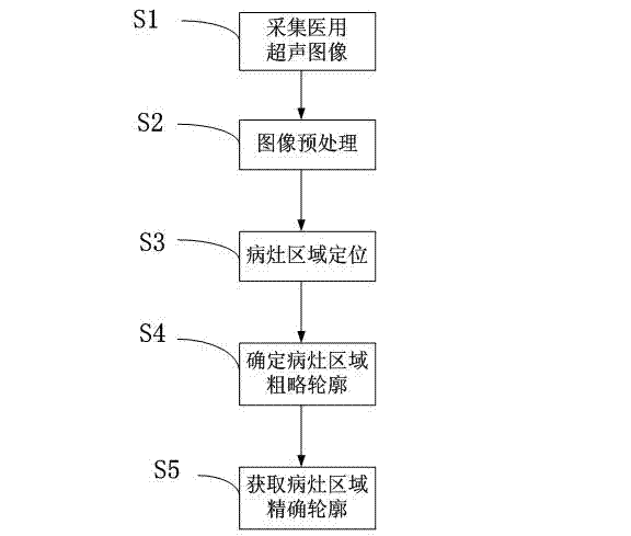
[0021] see figure 1 , figure 1 It is a schematic flowchart of the method for processing ultrasonic images of space-occupying lesions in the present invention. As shown in the figure, the technical solution of the present invention is to preprocess the collected ultrasound images, including removing text information around the images, filtering, border enhancement, determining effective information areas, etc.; The rough contour of the lesion area is extracted using the rough contour as the initial contour of the active contour model algorithm. The process includes the following steps: S1, collecting medical ultrasound images; S2, performing preprocessing on the medical ultrasound...


PUM


Abstract

The invention discloses a processing method for space-occupying lesion ultrasonic images. The processing method preprocesses the acquired ultrasonic image, comprising the following steps: the removal of the text information around the image, filtration, edge enhancement, the determination of an effective information region and the like; automatic location of a lesion region; determination of the rough contour of the space-occupying lesion, and extraction of the precise contour of the lesion region with the rough contour as an initial contour for an active contour model algorithm. The processing method realizes the functions of automatically segmenting the space-occupying lesion ultrasonic image and automatically extracting the region of interest in order to automatically diagnose the space-occupying lesion, thus enhancing the objectivity and accuracy of clinical diagnosis, and therefore the processing method effectively assists the diagnosis of space-occupying lesions.

Description

technical field [0001] The invention relates to the field of ultrasonic images, in particular to a processing technology for ultrasonic images of space-occupying lesions. Background technique [0002] Cancer is a general term for a large class of malignant tumors. In the early stage of cancer, there are usually few symptoms, and a series of symptoms and signs will gradually appear after it develops to a certain stage. At this time, it is often too late to take treatment measures. Therefore, early diagnosis and early treatment of cancer are particularly critical. At present, the main methods for early diagnosis of cancer include alpha-fetoprotein detection, ferritin detection, ultrasonography, CT examination, magnetic resonance imaging, selective contrast examination, etc. Among them, ultrasonography has the characteristics of non-invasive, radiation-free, low-cost, real-time, repeatable and easy to operate, and has become one of the main methods for early diagnosis of canc...

Claims


Application Information

Patent Timeline
Patent Type & Authority Patents(China)
IPC IPC(8): A61B8/08G06T7/00
Inventor 汪天富王毅陈思平和小孔
Owner SHENZHEN UNIV
Why Eureka
Try Eureka