Microfluidic imaging cytometry
A technology of microfluidic system and microfluidic chip, which is applied in the field of microfluidic system and can solve problems such as no exploration platform
- Summary
- Abstract
- Description
- Claims
- Application Information
AI Technical Summary
Problems solved by technology
Method used
Image

Examples
preparation example Construction
[0098] 1. Cell culture sample preparation, including cell loading, culturing cells in an incubator, and media exchange for maintaining cells.
[0099] 2. Immunocytochemistry: including cell fixation, permeabilization and immunostaining.
[0100] According to some embodiments of the present invention, microfluidic system 100 may provide a system for (i) large-scale cell culture for high-throughput screening, (ii) microfluidic cell analysis for precise quantification of single-cell biomolecules , (iii) signaling pathway network analysis associated with cancer diagnosis and therapeutic classification, (iv) dynamic protein quantification as an alternative to Western blotting, and other broader applications for quantitative analysis of proteomics in cells.
[0101] Potential applications for the microfluidic system 100 may include, but are not limited to:
[0102] 1. Alternative techniques to flow cytometry, which may include the following advantages: (i) low cost, (ii) less patie...
Embodiment A
[0119] Example A: Determination Example
[0120] 1) Sample preparation, including cell loading, culturing cells in an incubator, and media exchange for maintaining cells.
[0121] 2) Immunocytochemistry: including cell fixation, permeabilization and immunostaining.
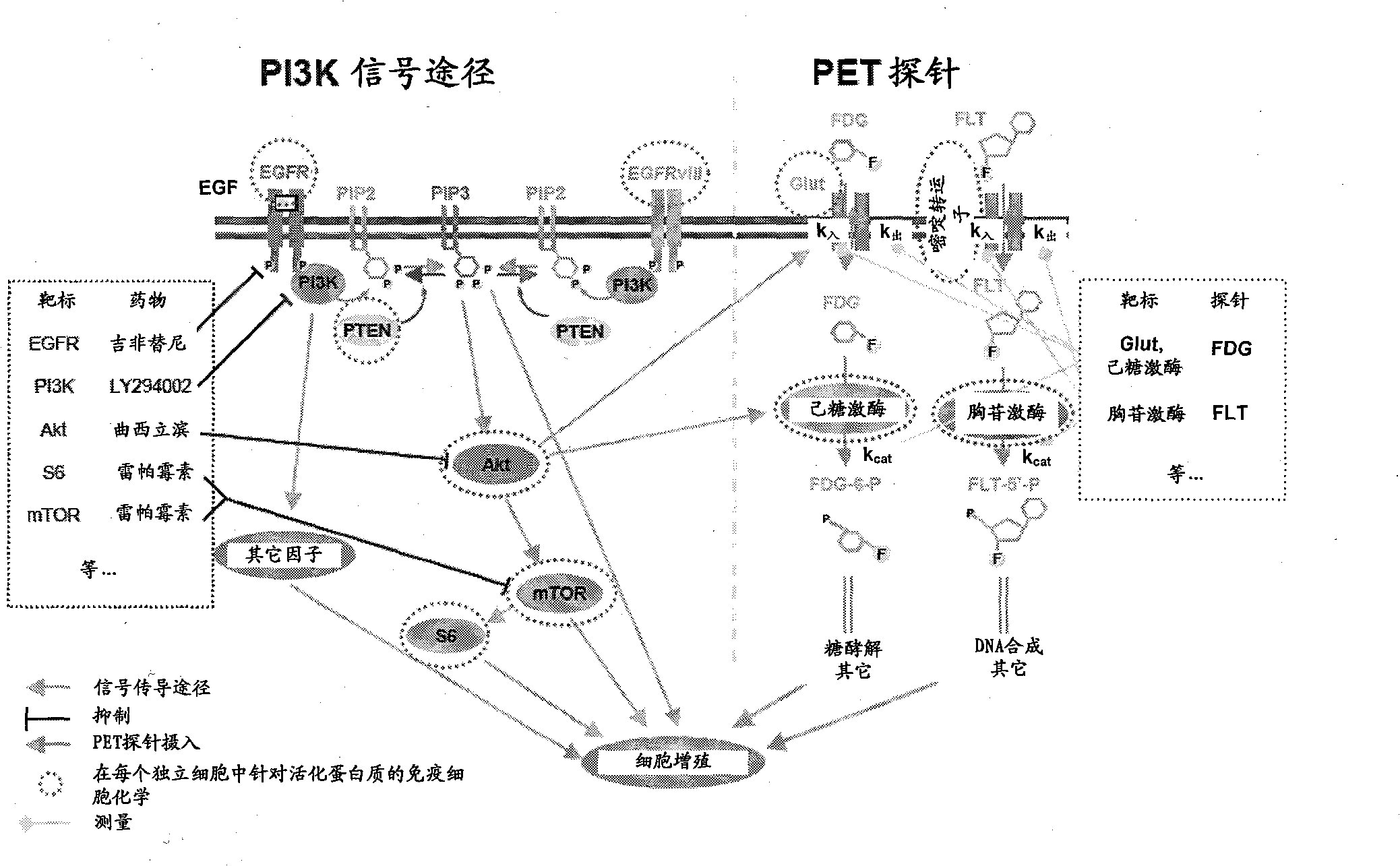
[0122] Can measure various signaling nodes (including EGFR, EGFvIII, PTEN, pAkt, pmTOR, pS6) involved in the PI3K-Akt-mTOR signaling network in the glioblastoma system and the proliferation marker Ki67.
[0123] Can measure various signaling nodes (including EGFR, ErbB2, PTEN, pAkt, pmTOR, pS6) and proliferation marker Ki67 involved in the PI3K-Akt-mTOR signaling network in the breast cancer system.
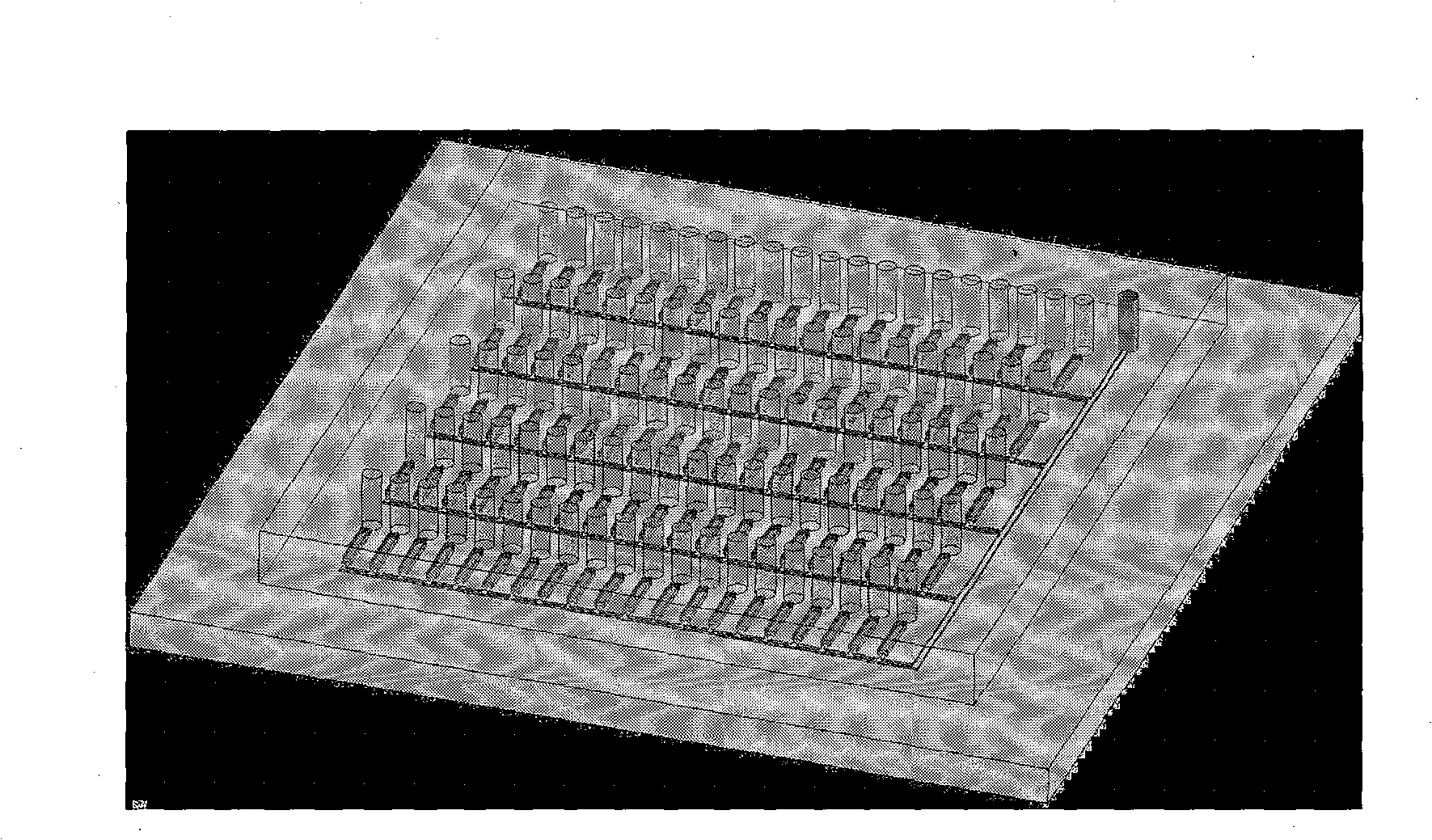
[0124] The growth curve of live cells can be monitored as follows:
[0125] The cell culture / assay chip shown in Figure 8 consists of two types of microchannels responsible for (i) parallel manipulation of 72 cell cultures and assays and (ii) wetting of adjacent cell culture chambers (to prevent media evaporation)....
Embodiment B
[0127] Example B: Data Collection
[0128] The data obtained by the methods of some embodiments of the invention may take the form of, for example, two-dimensional or three-dimensional dot plots (X-axis - strength of one signaling node (EGFRvIII in this case), Y-axis - another signaling Intensity of the node (DAPI in this case), for a 3D dot plot, Z-axis - intensity of the third signaling node))( Figure 9 ). In another embodiment ( Figure 10 ), the data can be in the form of a histogram (X-axis - strength of one signaling node (EGFRvIII in this case) and Y-axis - cell number).
[0129] Procedures for culturing glioblastoma cells in a chip
[0130] Material:
[0131] 24-channel poly-L-lysine coated cell culture chip
[0132] Cell culture medium: 500mL Dulbecco's Modified Eagle medium
[0133] 50mL fetal bovine serum
[0134] 5mL penicillin-streptomycin / L-glutamine
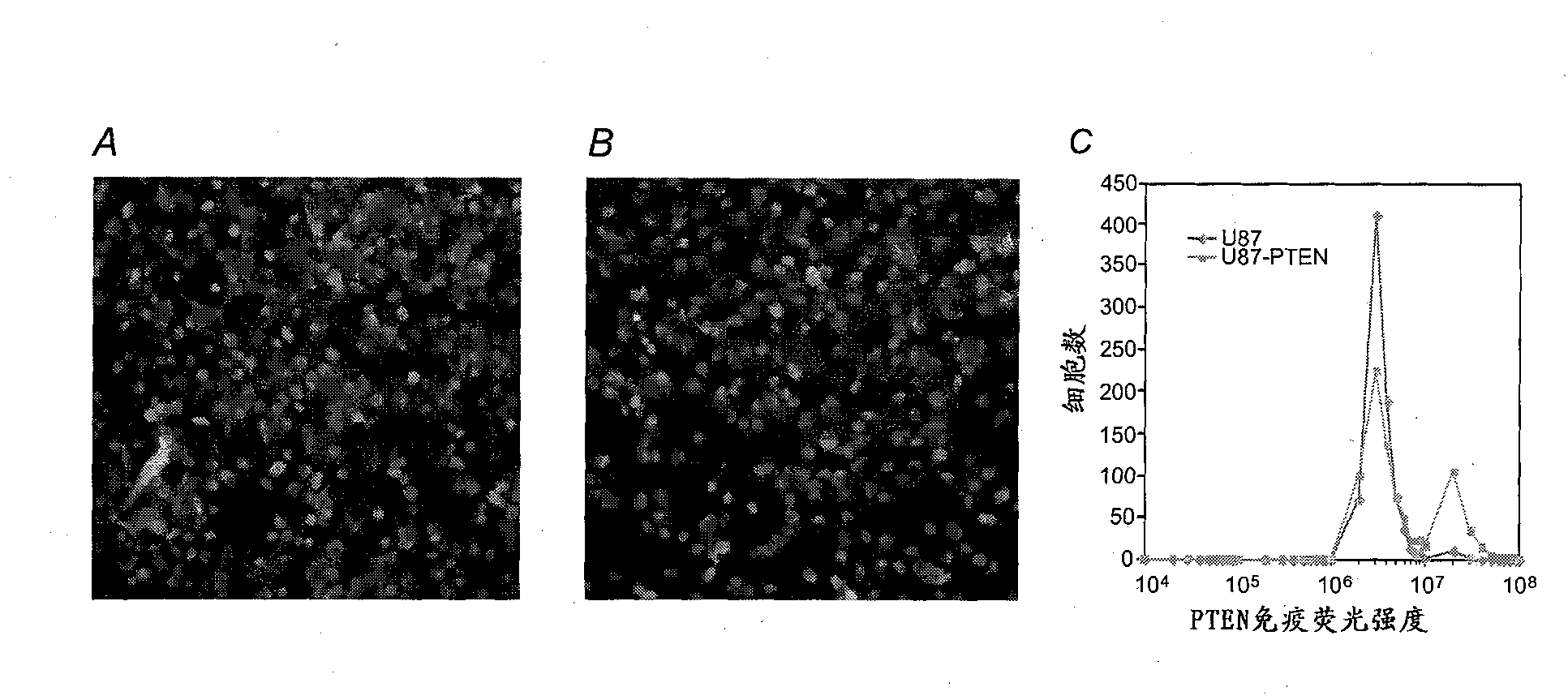
[0135] Cell lines and isogenetic cells:
[0136] U87
[0137] U87-PTEN
[0138] U87-EGFR
[0139] U...
PUM
| Property | Measurement | Unit |
|---|---|---|
| volume | aaaaa | aaaaa |
| diameter | aaaaa | aaaaa |
| Sensitivity | aaaaa | aaaaa |
Abstract
Description
Claims
Application Information
 - R&D Engineer
- R&D Manager
- IP Professional
- Industry Leading Data Capabilities
- Powerful AI technology
- Patent DNA Extraction
Browse by: Latest US Patents, China's latest patents, Technical Efficacy Thesaurus, Application Domain, Technology Topic.
© 2024 PatSnap. All rights reserved.Legal|Privacy policy|Modern Slavery Act Transparency Statement|Sitemap