Digital sternum heart calcification image intensification method

An image enhancement and chest X-ray technology, applied in the field of cardiac calcification image enhancement technology, can solve the problems of low contrast and difficult identification of cardiac calcification images, and achieve the effects of reducing labor intensity, improving reading efficiency, and shortening reading time.

Active Publication Date: 2009-05-13
XI AN JIAOTONG UNIV
View PDF0 Cites 3 Cited by
  • Summary
  • Abstract
  • Description
  • Claims
  • Application Information

AI Technical Summary

Problems solved by technology

[0007] In order to solve the problem of low contrast and difficulty in identifying cardiac calcification images in existing digital chest radiographs, the present invention proposes a simple and practical method that can effectively enhance cardiac calcification images to meet the requirements of digital chest radiograph operators and readers. The need for image enhancement of cardiac calcification

Method used


Image

  • Digital sternum heart calcification image intensification method
  • Digital sternum heart calcification image intensification method
  • Digital sternum heart calcification image intensification method

Examples


Embodiment Construction

[0041] The present invention will be further described in detail below in conjunction with the accompanying drawings and embodiments.

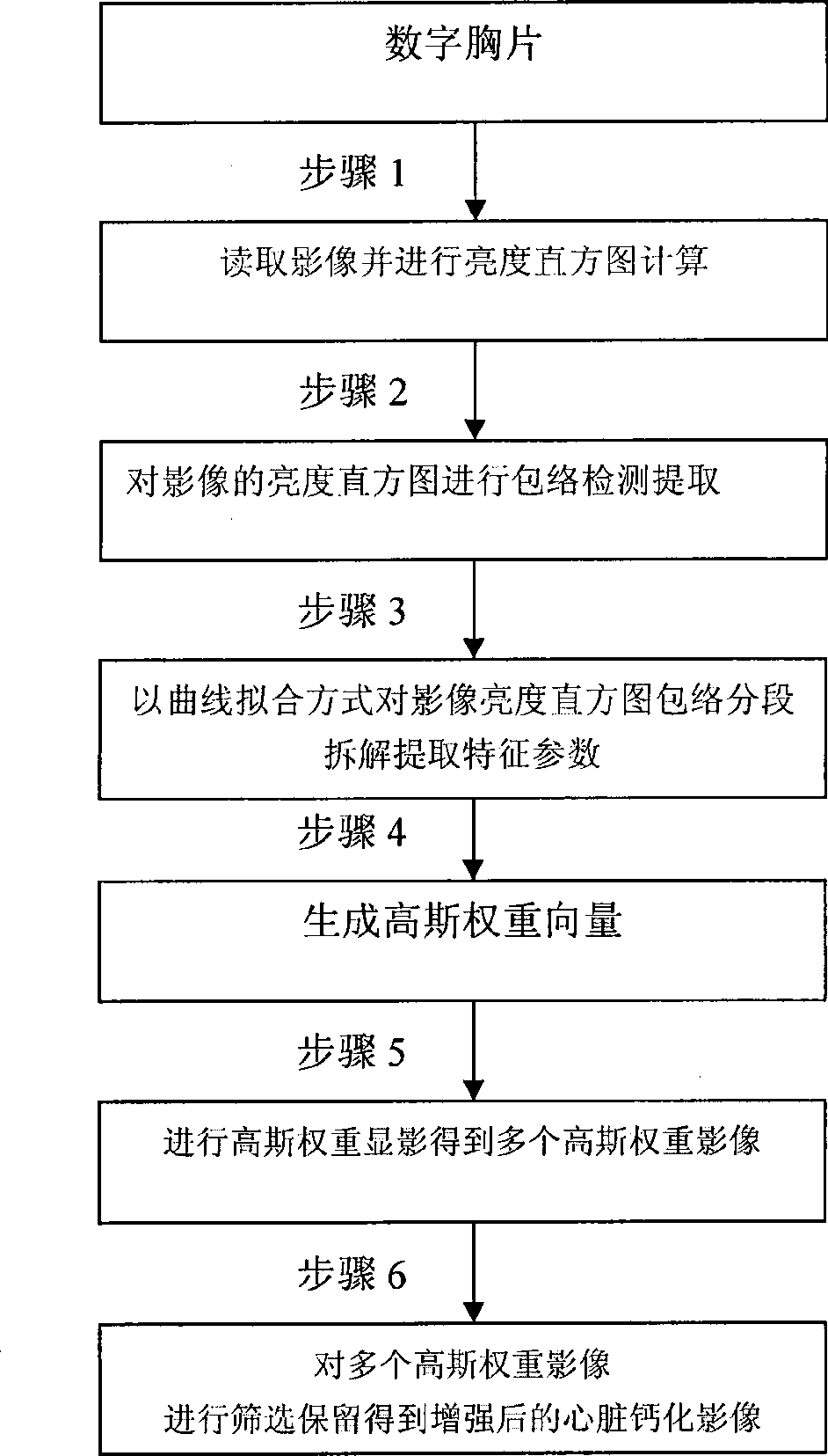
[0042] like figure 2 Shown, a kind of digital chest X-ray cardiac calcification image enhancement method comprises the following steps:

[0043]Step 1, read the original digital chest X-ray image, the digital chest X-ray image includes CR (Computed Radiography), DR (Digital Radiography), DES (Dual Energy Subtraction), DEDR (Dual Energy Digital Radiography), MEDR (Multi-Energy Digital Radiography) Chest images of the human body and other mammals obtained by other methods. The conventional brightness histogram calculation is performed on the digital chest radiograph image.

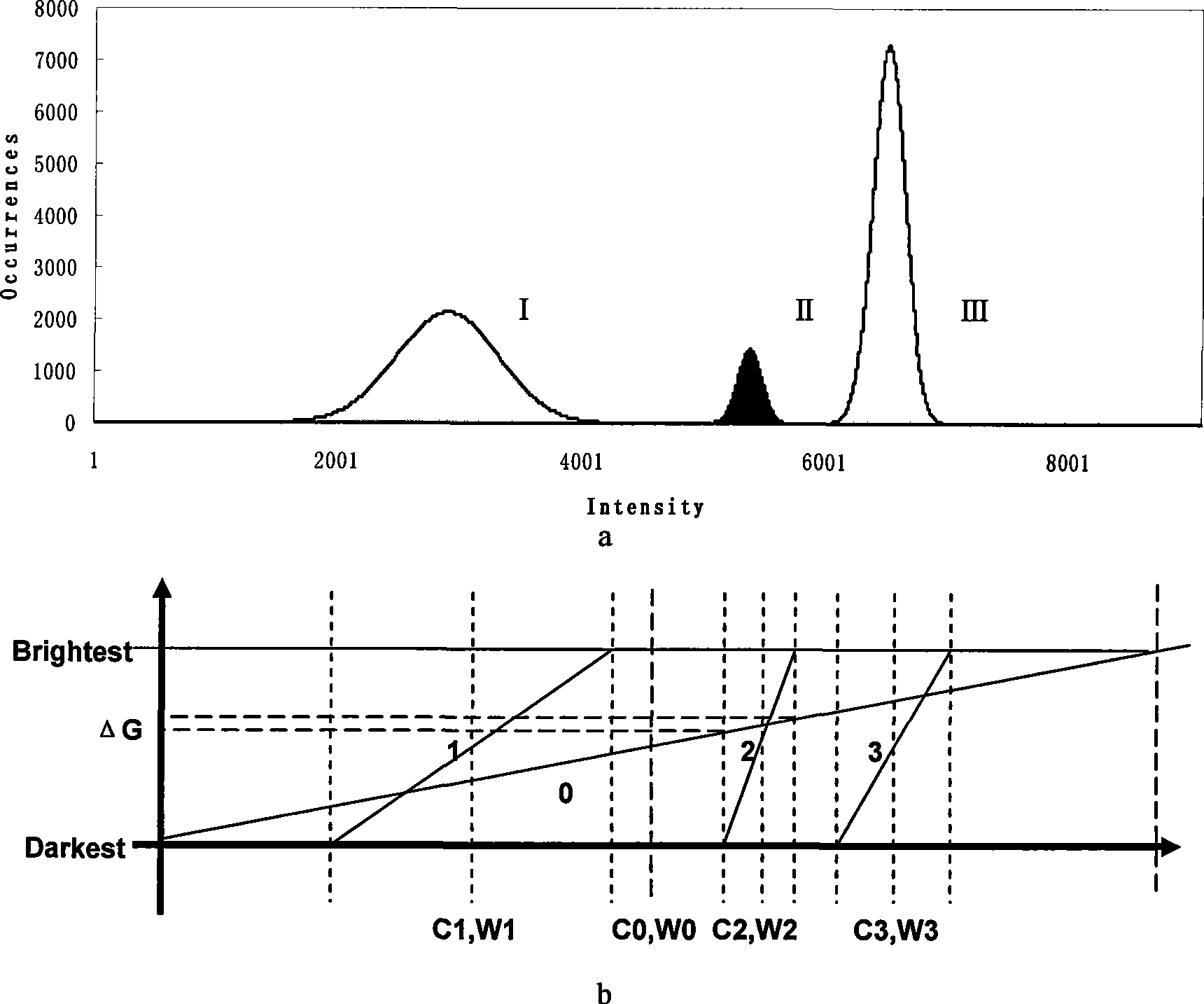
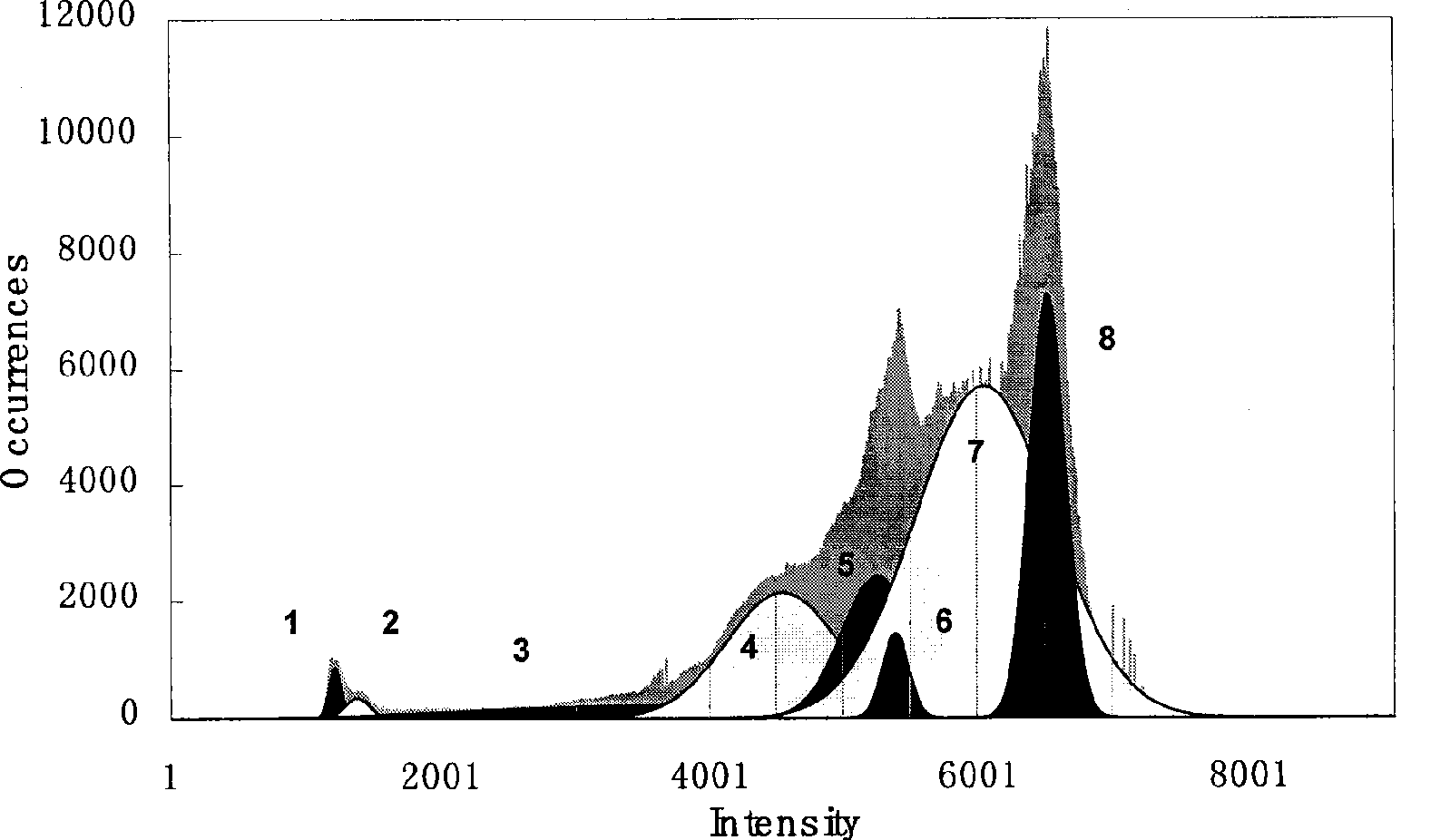
[0044] Step 2, carry out envelope detection according to formula (1), (2) description of the brightness histogram of this digital chest radiograph image:

[0045] SH ( i ) = ...


PUM


Abstract

The invention discloses an enhancement method of a cardiac calcification image by a digital chest radiograph. The method comprises the following steps: disassembling a brightness histogram in a segmented manner by a Gaussian function; fitting the brightness histogram of the digital chest radiograph by a series of Gaussian functions to directly acquire characteristic parameters of the Gaussian functions; and realizing the enhancement of the cardiac calcification image by respectively performing weighted development on different Gaussian functions. In the method, the characteristic parameters of various Gaussian functions are directly determined by computing a fitting curve and an enveloped non-linear least square, which replaces a complex window width and window level self-adaptive adjustment process which is used in the prior art. The comparison and screening of a plurality of Gaussian weighted projection images replace the adjustment process of window parameters in the window technology, and a fine adjustment of the window width and the window level within a large range is simplified into the comparison and the screening of 3-8 weighted images, which causes the method to lower the labor intensity of a radiograph reader, reduce the radiograph reading time and improve the radiograph reading efficiency.

Description

technical field [0001] The invention relates to the field of visual analysis of medical images, and more particularly relates to an improved cardiac calcification image enhancement technology in digital chest radiographs. Background technique [0002] Since the German physicist Roentgen discovered X-rays in 1895, X-ray imaging technology has been widely used in various fields of medicine. Digital chest X-ray is a digital chest X-ray imaging method developed in recent years and based on ordinary X-ray chest X-ray technology. [0003] In view of the different contrast ratios of various tissues in digital chest films and the wide dynamic range of X-ray image brightness, in order to better display various tissue structures, the field of medical image visualization analysis has gradually formed a window display image centered on window width and window level adjustment. Enhancement technology, that is, by continuously adjusting the center position of the display window, that is,...

Claims


Application Information

Patent Timeline
IPC IPC(8): G06T5/00G06T5/40A61B6/02
Inventor 陈翔金捷李津孙卫新郭萍孔澍
Owner XI AN JIAOTONG UNIV
Why Eureka
Try Eureka