
Apparatus and method for displaying virtual endoscopy display

a virtual endoscope and display technology, applied in the field of apparatus and methods for displaying a three-dimensional virtual endoscope image, can solve the problems of difficult operation of conventional virtual endoscopes, difficult operation of virtual cameras in the desired direction, and difficulty in expressing a three-dimensional correlation and detecting the position of parts, so as to facilitate the operation of virtual cameras and effectively show correlations

Inactive Publication Date: 2006-09-05
INFINITT HEALTHCARE CO LTD
View PDF16 Cites 45 Cited by
  • Summary
  • Abstract
  • Description
  • Claims
  • Application Information

AI Technical Summary

Benefits of technology

"The present invention provides an apparatus and method for displaying a three-dimensional virtual endoscopic image that effectively shows a correlation between reference images and the virtual endoscopic image. The virtual camera is intuitively displayed on the reference images for easy operation. The apparatus includes a volume data input unit, a two-dimensional reference image display control unit, a three-dimensional reference image display control unit, a virtual endoscopic image display control unit, a user interface unit, and a controller. The method includes inputting information on the virtual endoscopic image, detecting reference images and the virtual endoscopic image from the volume data, displaying the reference images and the virtual endoscopic image on one screen, and displaying virtual cameras on areas where the reference images are displayed. The technical effect of the invention is to provide a more realistic and effective display of a virtual endoscopic image."

Problems solved by technology

However, a conventional virtual endoscope is difficult to operate due to inefficient user interface.
Accordingly, in the conventional virtual endoscopic method, a correlation between the reference image and the virtual endoscopic image is two-dimensionally shown, so it is difficult to express a three-dimensional correlation and detect the position of a part shown in the virtual endoscopic image or a relative relation with other structures.
Accordingly, it is difficult for a user to operate the virtual camera in a desired direction and recognize the direction of the virtual camera.

Method used


Image

  • Apparatus and method for displaying virtual endoscopy display
  • Apparatus and method for displaying virtual endoscopy display
  • Apparatus and method for displaying virtual endoscopy display

Examples


Embodiment Construction

[0021]Hereinafter, embodiments of the present invention will be described in detail with reference to the attached drawings.

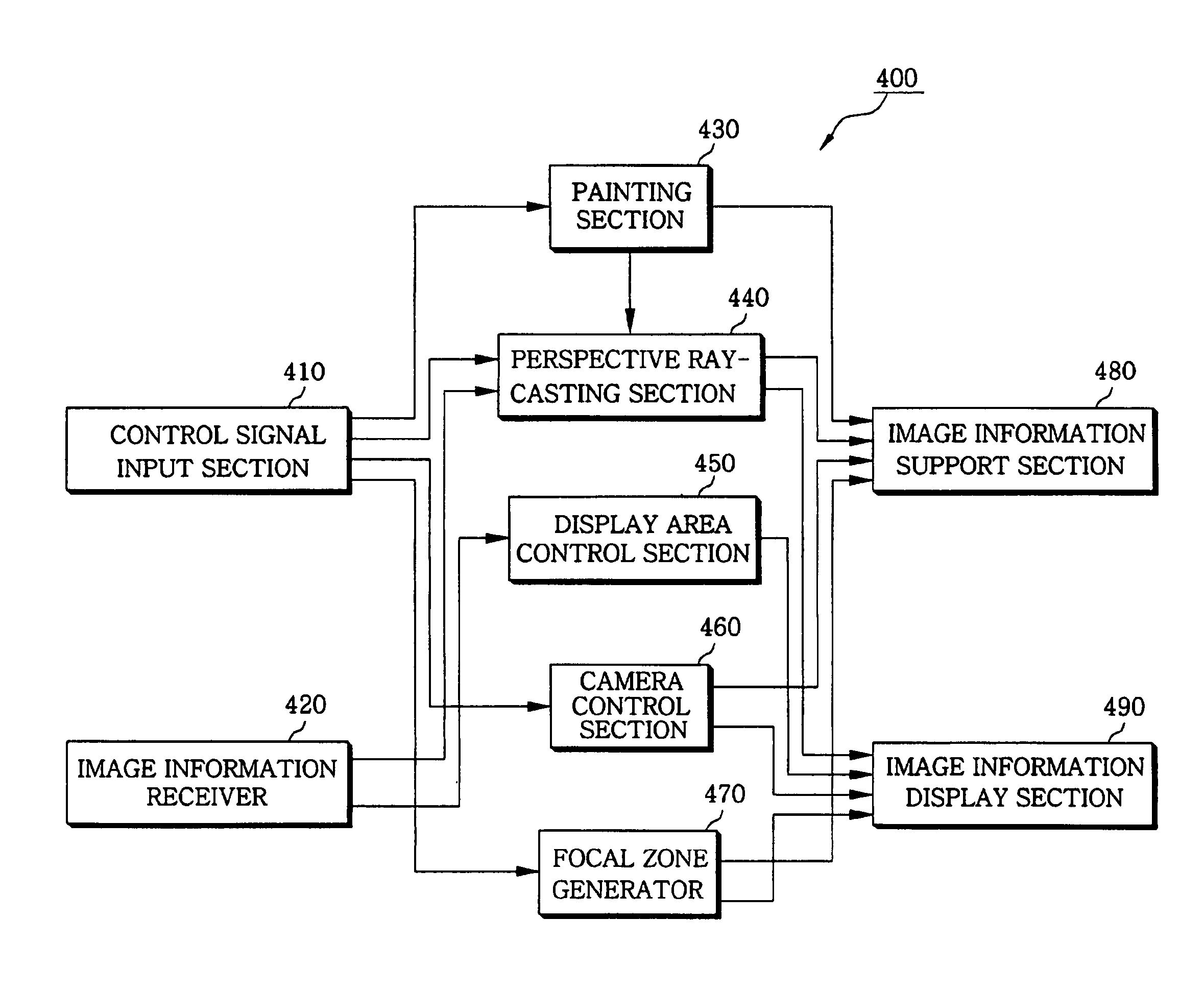
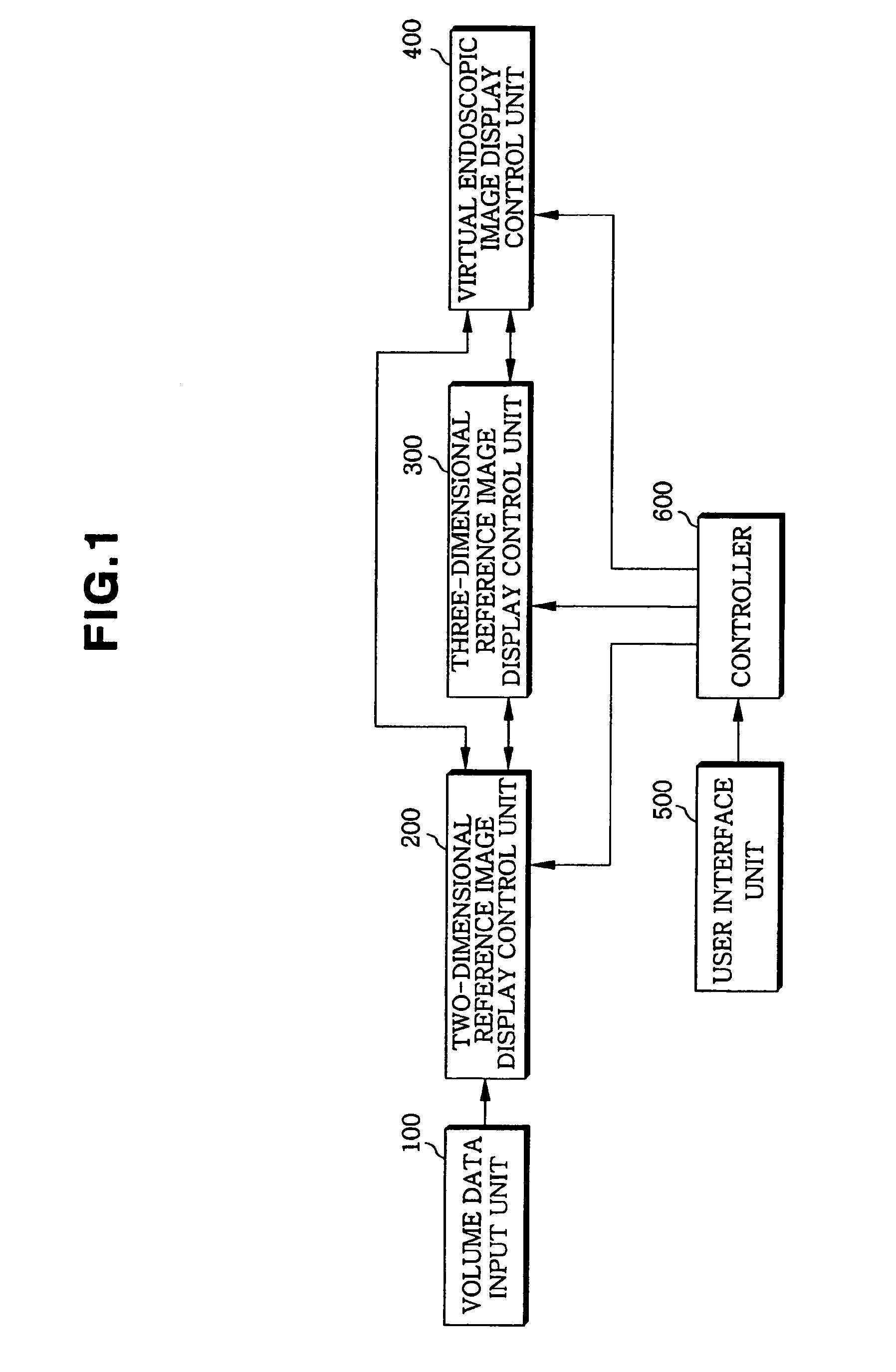
[0022]FIG. 1 is a schematic block diagram of an apparatus for displaying a three-dimensional virtual endoscopic image according to an embodiment of the present invention. Referring to FIG. 1, the apparatus for displaying a three-dimensional virtual endoscopic image includes volume data input unit 100, a two-dimensional reference image display control unit 200, a three-dimensional reference image display control unit 300, a virtual endoscopic image display control unit 400, a user interface unit 500, and a controller 600.

[0023]The volume data input unit 100 inputs information on a virtual endoscopic image in the form of volume data expressed as a function of three-dimensional position. For example, in a case where volume data, which is generated as the result of computed tomography (CT) scan or magnetic resonance imaging (MRI), is directly received from each dev...


PUM


Abstract

An apparatus and method for displaying a three-dimensional virtual endoscopic image are provided. In the method, information on a virtual endoscopic image is input in the form of volume data expressed as a function of three-dimensional position. A two-dimensional reference image, a three-dimensional reference image, and a virtual endoscopic image are detected from the volume data. The detected images are displayed on one screen. Virtual cameras are respectively displayed on areas, in which the two and three-dimensional reference images are respectively displayed. Here, a camera display sphere and a camera display circle are defined on the basis of a current position of each virtual camera. When information regarding to one image, among the two and three-dimensional reference images and the virtual endoscopic image on one screen, is changed by a user's operation, information regarding to the other images is changed based on the information changed by the user's operation.

Description

TECHNICAL FIELD[0001]The present invention relates to an apparatus and method for displaying a three-dimensional virtual endoscopic image, and more particularly, to an apparatus and method for displaying a three-dimensional virtual endoscopic image, through which a virtual endoscopic image is displayed along with two- and three-dimensional reference images, a virtual camera is intuitively displayed on the two- and three-dimensional reference images, and the images are controlled to be associated with one another.[0002]Generally, three-dimensional image processing systems are applied to various fields such as design and construction of structures, for example, cars and ships, having a three-dimensional shapes and medical imaging. In particular, applying three-dimensional image processing systems to the field of medical imaging is referred to as a “three-dimensional medical imaging”. Three-dimensional medical imaging is generating a three-dimensional image from a series of two-dimensi...

Claims


Application Information

Patent Timeline
Patent Type & Authority Patents(United States)
IPC IPC(8): G06T15/00G06K9/00G06T15/20G09G5/00A61B1/00G02B27/22G06T15/08
CPCG06T15/08G06T19/00G06T2219/028G06T2219/008G06T2210/41A61B1/00
Inventor KIM, BO-HYOUNGSHIN, YEONG-GILCHUNG, JIN-WOOK
Owner INFINITT HEALTHCARE CO LTD
Learn More