
Agents for treatment of glaucomatous retinopathy and optic neuropathy

a technology of optic nerve and glaucoma, applied in the field of retinopathy and optic neuropathy, can solve the problems of not addressing the protection or treatment of cannot be predicted from the biological responses of spinal cord tissues and brain tissues, and the application cited does not address the loss of retinal ganglion cells (rgc), so as to enhance the availability or the transport of nrf2. the effect of gene products and enhancing the availability of gene products

Inactive Publication Date: 2011-06-16
NOVARTIS AG
View PDF31 Cites 0 Cited by
  • Summary
  • Abstract
  • Description
  • Claims
  • Application Information

AI Technical Summary

Benefits of technology

[0021]According to the present invention, an agent having stimulatory activity for Nrf2 protein nuclear translocation and the subsequent increases in gene products that detoxify and eliminate cytotoxic metabolites provides a protective or therapeutic effect in delaying or preventing loss of retinal ganglion cells and glaucomatous damage to the optic nerve. As used herein “stimulatory activity for Nrf2 protein nuclear translocation” means an agent that enhances the availability or the transport of Nrf2 to the nucleus. Translocation of Nrf2 protein to the nucleus allows a subsequent increase in expression of gene products that detoxify and eliminate cytotoxic metabolites. The methods of the present invention provide a method of treatment for glaucomatous retinopathy and optic neuropathy in a subject comprising administering to the subject an effective amount of a composition comprising an agent having stimulatory activity for Nrf2 protein nuclear translocation, and an acceptable carrier. The subject may be at risk for developing glaucomatous retinopathy or optic neuropathy or may have symptoms of glaucomatous retinopathy or optic neuropathy.

Problems solved by technology

Nonetheless, the loss of visual field in glaucoma patients does not always correlate with IOP, and lowering IOP alone does not completely stop the disease process.
However, none of these proposed mechanisms is universally accepted by researchers in the field.
Further, biological responses of ocular tissues such as the retina to particular therapeutic agents cannot be predicted from the biological responses of spinal cord tissues and brain tissues.
The cited applications do not address protection or treatment for loss of retinal ganglion cells (RGC) and optic neuropathy in glaucoma.
There is no generally accepted anti-glaucoma therapeutic method to manage glaucomatous retinopathy and optic neuropathy.

Method used


Image

  • Agents for treatment of glaucomatous retinopathy and optic neuropathy

Examples


example 1

Agents Having Stimulatory Activity for Nrf2 Protein Nuclear Translocation

[0058]Vascular endothelial cells, such as bovine aortic endothelial cells (BAEC, VEC Technologies, Rensselaer, N.Y.), are used to determine those agents having stimulatory activity for Nrf2 protein nuclear translocation. For example, confluent monolayers of bovine aortic endothelial cells are exposed to candidate agents in Dulbecco's modified Eagle's medium with 1% fetal bovine serum for up to 24 hours. Cell lysates, cytosolic extracts, and nuclear extracts are prepared, and immunoblotting performed and quantified as described in Buckley, B. J., et al. (Biochem Biophys Res Commum, 307:973-979 (2003)). Agents that increase the amount of Nrf2 detected in the nuclear fraction as compared to control cells without agent are then tested for activity in a retinal ganglion cell toxicity assay as set forth in Example 2.

example 2

Protection of Rat Retinal Ganglion Cells by an Agent Having Stimulatory Activity for Nrf2 Protein Nuclear Translocation

[0059]Cultured rat neural retinal cells are combined with an agent that stimulates nuclear translocation of Nrf2 protein for 1 to 24 hours, then the combination is exposed to peroxide. Survival of the neural retinal cells as compared to a control culture without peroxide indicates that the agent provides protection from the oxidant.

[0060]Neonatal rat neural retinal cells are isolated and cultured as reported in Pang, I-H., et al, (Invest Ophthalmol V is Sci 40:1170-1176 (1999)). “Neural retinal” refers to the retina without the retinal pigment epithelium. Thus, the culture contains a mixed population of retinal cell types. Briefly, neonatal Sprague-Dawley rats 2-5 days old are anesthetized and a 2 mm midline opening is made in the scalp just caudal to the transverse sinus. Retinal ganglion cells are selectively retrograde labeled with a fluorescent dye, Di-I, (1,1′-...

example 3

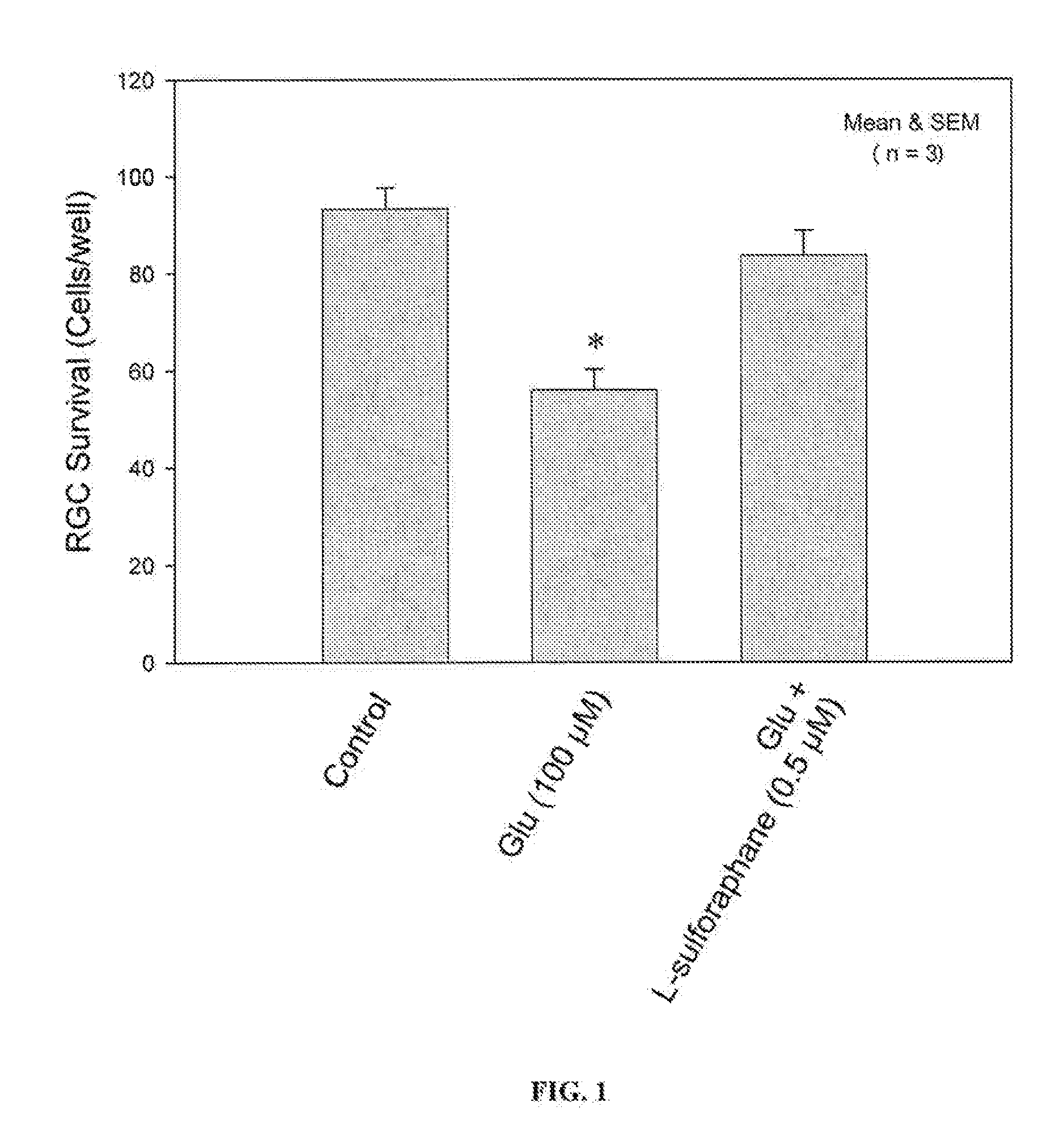
Protection of Rat Retinal Ganglion Cells from Glutamate-Induced Toxicity by Sulforaphane

[0063]Adult Sprague-Dawley rats were euthanized by CO2 asphyxiation. Their eyes were enucleated and placed in NEUROBASAL™ medium (Gibco, Gaithersburg, Md.). The retina from each eye was detached and isolated. Retinal cells were dissociated by combining up to 20 retinae with 5 mL of papain solution containing 10 mg papain, 2 mg DL-cysteine, and 2 mg bovine serum albumin in 5 ml of NEUROBASAL™ medium, for 25 min at 37° C., then washed 3 times with 5 mL RGC medium (NEUROBASAL™) medium supplemented as cited in Example 2 and with 1% fetal calf serum. Retinal pieces were triturated by passing through a fire-polished disposable pipet several times until cells were dispersed. The cell suspension was placed onto a poly-D-lysine- and laminin-coated 8-well chambered culture slide. Glutamate and glutamate with sulforaphane were added to assigned wells. The cells were then cultured at 95% air / 5% CO2 at 37° C....


PUM

PropertyMeasurementUnit
intraocular pressureaaaaaaaaaa
pressureaaaaaaaaaa
antioxidant responseaaaaaaaaaa

Abstract

Agents that stimulate nuclear translocation of Nrf2 protein and the subsequent increases in gene products that detoxify and eliminate cytotoxic metabolites are provided in a method for treating glaucomatous retinopathy or optic neuropathy. The structurally diverse agents that act on the Nrf2 / ARE pathway induce the expression of enzymes and proteins that possess chemically versatile cytoprotective properties and are a defense against toxic metabolites and xenobiotics. Agents include certain electrophiles and oxidants such as a Michael Addition acceptor, diphenol, thiocarbamate, quinone, 1,2-dithiole-3-thione, butylated hydroxyanisole, flavonoid, an isothiocyanate, 3,5-di-tert-butyl-4-hydroxytoluene, ethoxyquin, a coumarin, combinations thereof, or a pharmacologically active derivative or analog thereof.

Description

[0001]This application is a continuation of application Ser. No. 11 / 015,888 filed Dec. 17, 2004, which claims the benefit of U.S. Provisional Patent Application No. 60 / 531,770, filed Dec. 22, 2003, which is incorporated by reference in its entirety.FIELD OF THE INVENTION[0002]The present invention relates to the field of prophylactic agents and therapeutics for retinopathy and optic neuropathy related to glaucoma.BACKGROUND OF THE INVENTION[0003]Glaucoma is a heterogeneous group of diseases that have a similar set of clinical features including optic nerve damage and selective apoptotic death of retinal ganglion cells (RGC), which leads to a progressive loss of visual field and blindness. An abnormal increase in intraocular pressure (IOP) is associated with most forms of glaucoma. The only available treatment is to lower IOP either by medication or surgery. Lowering IOP is effective in slowing the development of certain types of glaucoma and delaying its damaging effects. Nonetheles...

Claims


Application Information

Patent Timeline
Patent Type & Authority Applications(United States)
IPC IPC(8): A61K31/497A61K31/352A61K31/085A61K31/47A61K31/37A61K31/05A61K31/27A61K31/145A61K31/122A61P27/02A61P27/06A61K31/325A61K31/353A61K31/366A61K31/385A61K31/7048A61K45/00
CPCA61K9/0014A61K9/0024A61K9/0043A61K9/0048A61K45/06A61K31/353A61K31/366A61K31/385A61K31/7048A61K31/325A61P27/00A61P27/02A61P27/06A61P43/00
Inventor LANDERS, ROBERT A.PANG, IOK-HOU
Owner NOVARTIS AG
Learn More