
Line structure detection and analysis for mammography cad

a line structure and mammography technology, applied in the field of computer-aided cancer detection, can solve the problems of significant differences between the different approaches, high rate of unnecessary biopsies, and limited efficacy and efficiency of the above-mentioned methods

Inactive Publication Date: 2009-02-12
CARESTREAM HEALTH INC
View PDF7 Cites 11 Cited by
  • Summary
  • Abstract
  • Description
  • Claims
  • Application Information

AI Technical Summary

Problems solved by technology

Distinguishing between malignant and benign clusters is difficult and time-consuming for radiologists, which may lead to a high rate of unnecessary biopsies that may be avoided or at least minimized if a computer aided detection (CAD) mammography system is employed in diagnosis.
Results obtained using synthetic images suggest significant differences between the different approaches.
The aforementioned methods demonstrate limited success with regard to efficacy and efficiency because of the enormous variations of curvilinear structures to be detected in terms of shape geometric, topological properties and pixel luminance properties.

Method used


Image

  • Line structure detection and analysis for mammography cad
  • Line structure detection and analysis for mammography cad
  • Line structure detection and analysis for mammography cad

Examples


Embodiment Construction

[0021]In one embodiment of the method of image linear structure detection of the present invention, the mammographic image is a digitized X-ray film mammogram in the present invention; the mammographic image is a digital mammogram captured with a computerized radiography system in the present invention; the mammographic image is a digital mammogram captured with a digital radiography system in the present invention.

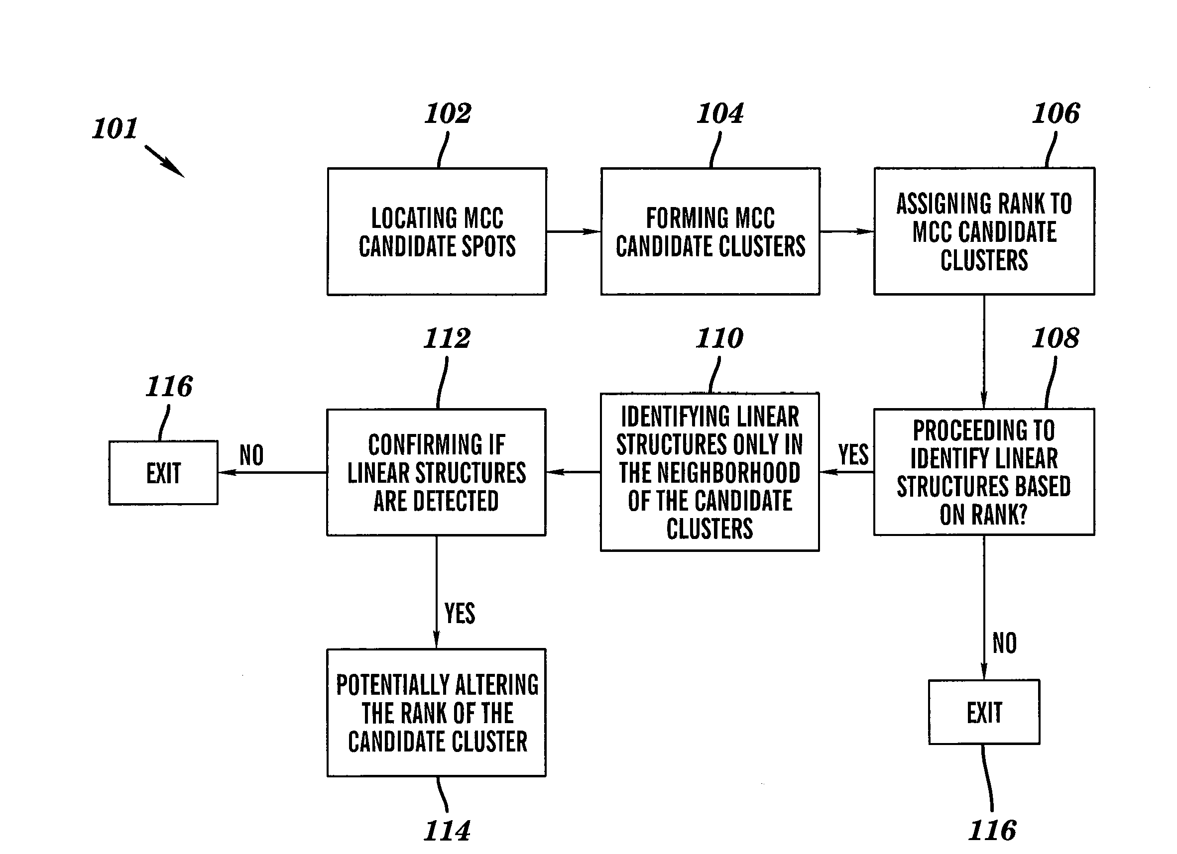
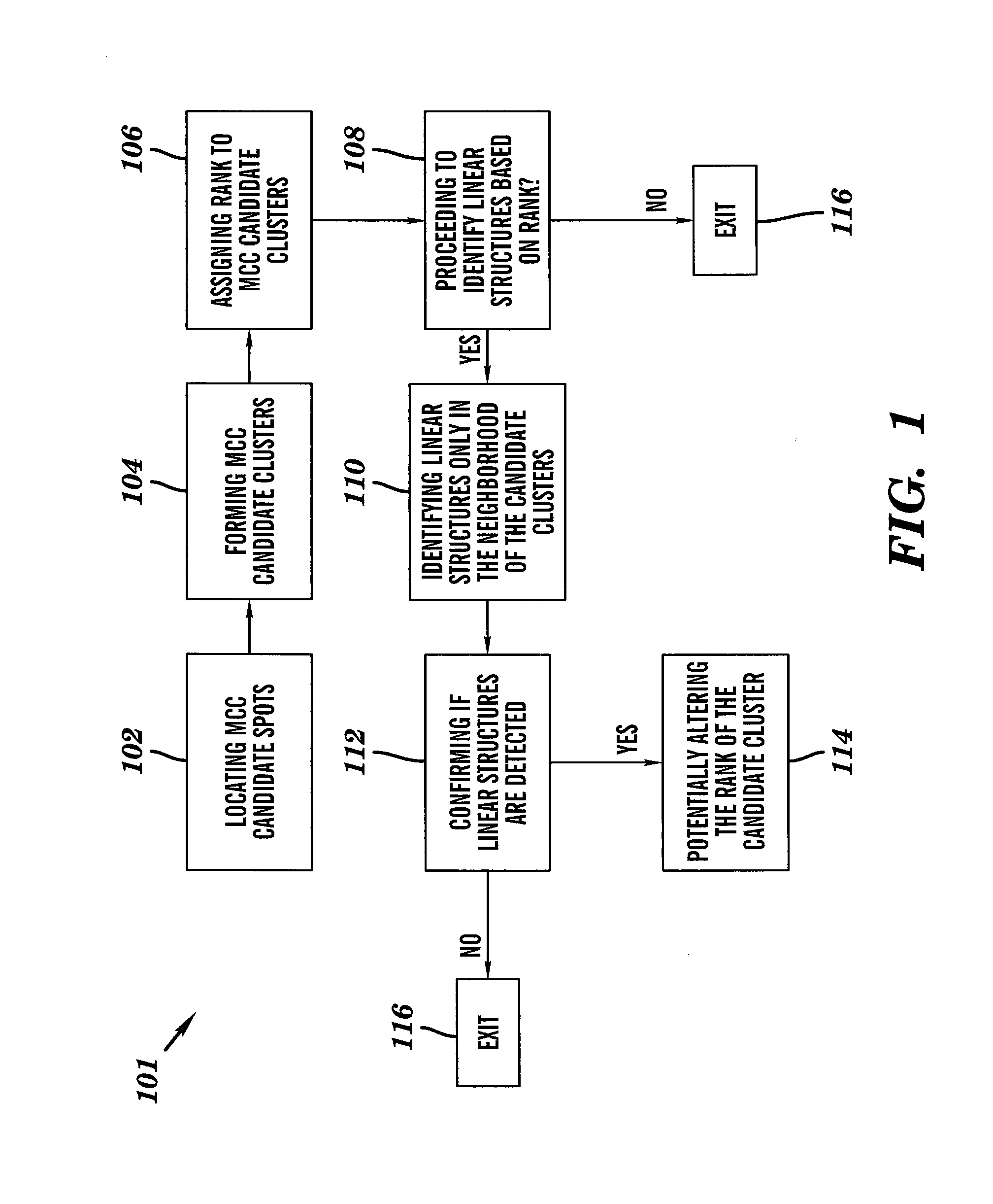
[0022]The step of locating microcalcification candidate spots in a mammographic image consists of a plurality of image processing and computer vision procedures that find clusters of connected pixels that present characteristics which are similar to that of microcalcification in mammogram.

[0023]The step of forming candidate clusters each of which has a plurality of microcalcification candidate spots groups a plurality of microcalcification candidate spots (mammogram image pixels) that are close to each other within a certain distance into a cluster. For each cluster, atta...


PUM


Abstract

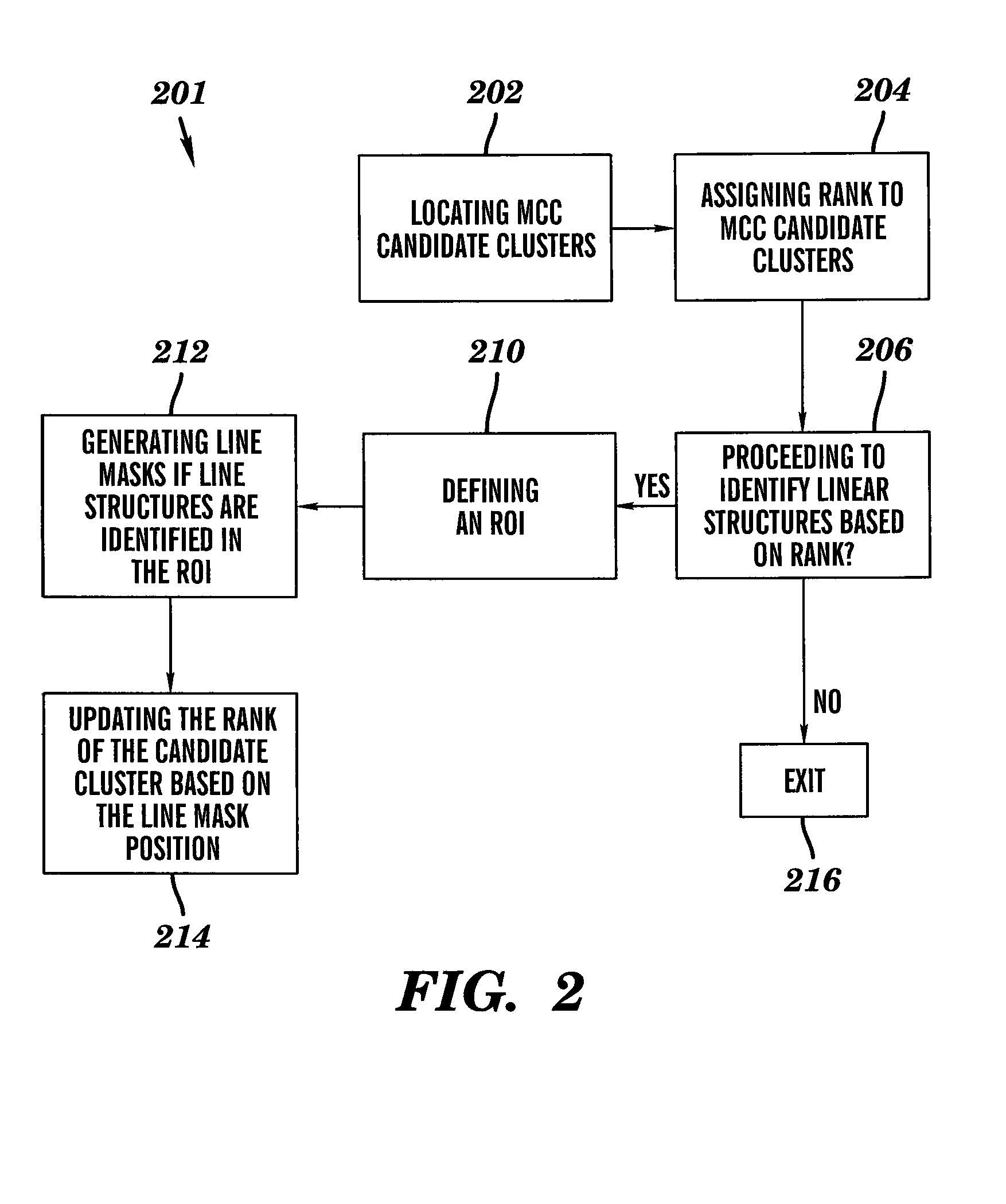
A method for image linear structure detection in medical imaging. The method includes locating microcalcification (mcc) candidate spots in a mammographic image; forming candidate clusters; assigning ranks to the candidate clusters; identifying linear structures in the neighborhood where the candidate clusters reside; and altering the ranks of the candidate clusters for which linear structures have been identified in the neighborhood.

Description

CROSS REFERENCE TO RELATED APPLICATIONS[0001]Reference is made to commonly assigned application U.S. Serial No. (Carestream Health, Inc. Docket No. 94203), entitled “LINEAR STRUCTURE VERIFICATION IN MEDICAL APPLICATIONS”, and filed on common date herewith in the names of Chen et al., and which is assigned to the assignee of this application.FIELD OF THE INVENTION[0002]The invention relates generally to computer aided cancer detection, and in particular, to mammography CAD.BACKGROUND OF THE INVENTION[0003]Digital or film-based mammography is widely employed for breast screening to reduce the cancer mortality among women. A sizable percentage of abnormality in mammograms are comprised of microcalcifications (mcc) that are deposits of calcium in breast carcinoma. A given cluster of microcalcifications might be associated with a malignant or benign case. Distinguishing between malignant and benign clusters is difficult and time-consuming for radiologists, which may lead to a high rate o...

Claims


Application Information

Patent Timeline
Patent Type & Authority Applications(United States)
IPC IPC(8): G06K9/00
CPCG06T7/0012G06T2207/10116G06T2207/30068
Inventor CHEN, SHOUPUZHANG, DAOXIAN HEIDISUN, YAJIECHU, YONGRAY, LAWRENCE A.ZHENG, YANGSHEN, YUE
Owner CARESTREAM HEALTH INC
Learn More