Analysis of P. aeruginosa infection in patients

a technology for detecting p. aeruginosa infection and bacterial burden in patients, which is applied in the direction of amide active ingredients, aerosol delivery, respiratory organ evaluation, etc., can solve the problems of increasing inflammation and lung damage in patients, no current method to rapidly determine, and cf patients, etc., and achieves the effects of not cost-effective, laborious, and time-consuming

Active Publication Date: 2008-12-11
STC UNM
View PDF4 Cites 15 Cited by
  • Summary
  • Abstract
  • Description
  • Claims
  • Application Information

AI Technical Summary

Problems solved by technology

However, there is no currently available method to rapidly determine whether a CF patient is actively infected with P. aeruginosa or to assess whole-lung bacterial levels.
Mucoid conversion is concomitant with the establishment of chronic bacterial colonization and increases inflammation and lung damage, while causing pulmonary function to decline.
Accordingly, mucoid conversion results in a poor prognosis for CF patients to heal Rapidly knowing if mucoid conversion has occurred, and the extent of mucoid pseudomonas burden would greatly improve therapies.
However, there is no currently available method to rapidly determine whether a CF patient is infected, or colonized with mucoid R aeruginosa or to assess whole-lung mucoid bacterial levels.
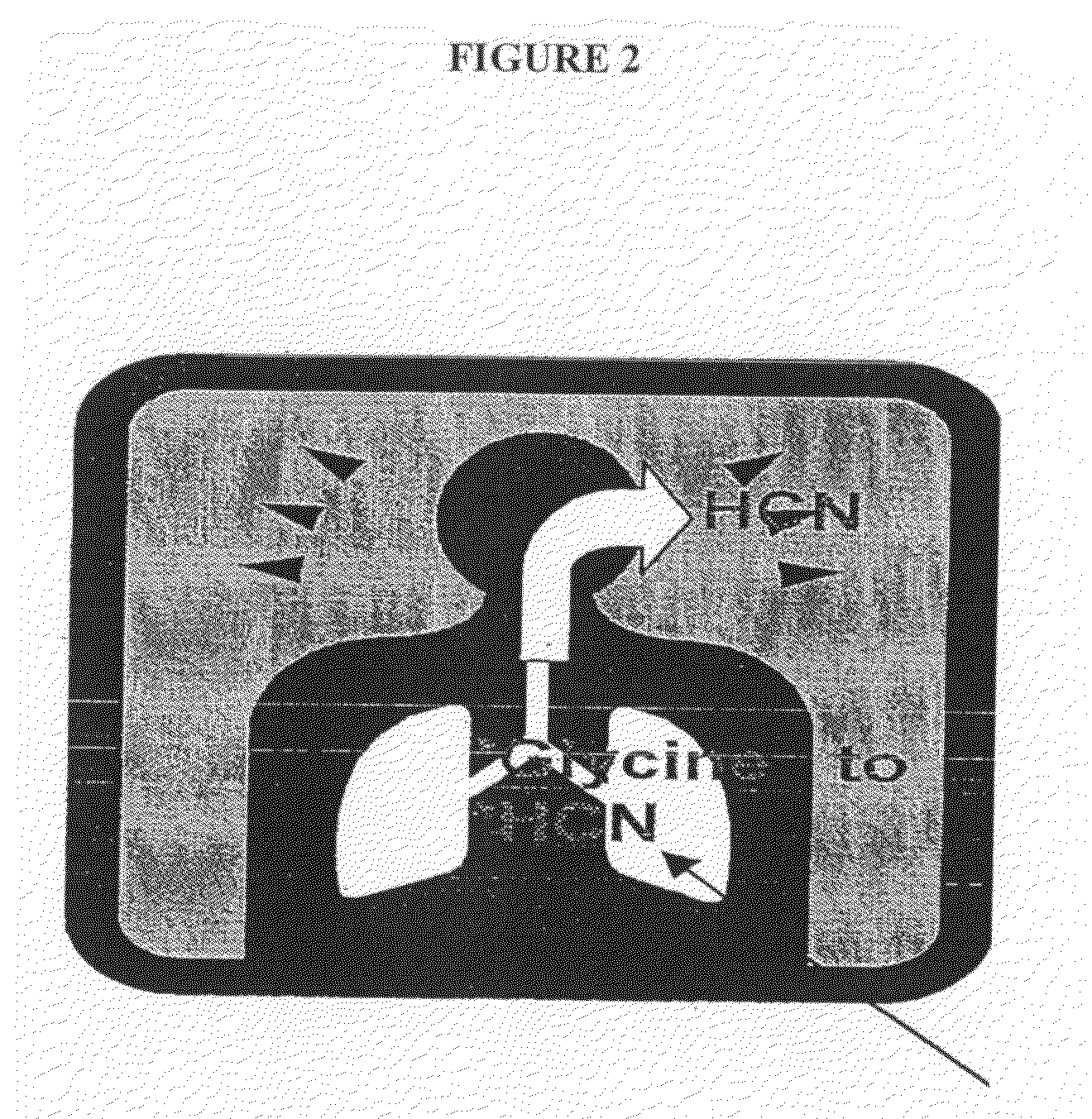
P. aeruginosa also expresses urease1-4, has the urease urea-ureG gene cluster whose expression is modifiable (Deretic & Timmins, unpublished microarray data), and consequently, is susceptible to detection by breath tests if the appropriate technology is developed and applied.

Method used


Image

  • Analysis of P. aeruginosa infection in patients
  • Analysis of P. aeruginosa infection in patients
  • Analysis of P. aeruginosa infection in patients

Examples


example 1

Inhaled Administration of Labeled Tracer

[0105]A. A first embodiment is to administer 13C-urea by inhalation (such as a dry powder inhaler as described above) in an amount of 0.1 to 10 mg, and the ratio of 13CO2 to 12CO2 in exhaled breath sample 1 to 60 minutes afterwards (to allow for conversion) is determined by mass spectrometry or laser spectroscopy. The ratio of 13CO2 to 12CO2 is compared to that of a breath sample obtained before compound inhalation, and an increase in this ratio is indicative of P. aeruginosa infection.

[0106]B. A second embodiment relates to replace the use of urea with the use of 15N-glycine as in the example A above, with analysis of the amount of HCN (labeled with 15N) compared to HCN in a sample prior to glycine administration in an exhaled breath sample 1 to 60 minutes after administration is determined by mass spectrometry or laser spectroscopy. The ratio of 15N to 14N is compared to that of a breath sample obtained before glycine inhalation, and an incr...

example 2

Oral Administration of Labeled Tracer

1) Oral Administration of Labeled Tracer

[0110]A.) One approach is to administer 13C-urea by in an oral dosage in an amount of about 0.1 to 100 mg, and the ratio of 13CO2 to 12CO2 in exhaled breath sample 1 to 180 minutes (also within the range of about 15-30 minutes to 60 minutes) after administration is determined by mass spectrometry or laser spectroscopy. The ratio of 13CO2 to 12CO2 is compared to that of a breath sample obtained before compound administration, and an increase in this ratio is indicative of P. aeruginosa infection. It may be desirable to preclude the potential for H. pylori interference with this assay by either enterically coating the urea capsule / tablet so that labeled urea does not come into contact with H. pylori and / or administration of known inhibitors of H. pylori urease such as bismuth salts, as otherwise described herein.

[0111]B) Another approach is to replace use of urea with the use of 15N-glycine with analysis of t...


PUM

PropertyMeasurementUnit
weightaaaaaaaaaa
massaaaaaaaaaa
timeaaaaaaaaaa

Abstract

The present invention relates to methods for detecting P aeruginosa infection and bacterial burden in the lungs of patients who are at risk for P. aeruginosa infections, especially including patients with Cystic Fibrosis (CF). The present method provides numerous tests (breath, blood, urine) which are readily administered to a patient that will sensitively and specifically detect the presence and extent of lung infection P. aeruginosa (both mucoid and non-mucoid), and allow monitoring of bacterial load as a parameter in monitoring treatment.

Description

RELATED APPLICATIONS AND FEDERALLY SPONSORED RESEARCH[0001]The present invention was made with government support under Grant Nos. A1 050825 and AI 31139 awarded by the National Institutes of Health. Consequently, the Government retains rights in this invention.[0002]The present application claims priority from U.S. provisional applications serial number US60 / 926,191, filed Apr. 25, 2007, serial number US60 / 927,670, filed May 4, 2007 and serial number US60 / 958,615, filed Jul. 6, 2007, each of which applications is incorporated by reference herein in its entirety.FIELD OF THE INVENTION[0003]The present invention relates to methods for detecting P aeruginosa infection and bacterial burden in the lungs of patients who are at risk for P. aeruginosa infections, especially including patients with Cystic Fibrosis (CF). The present method provides numerous tests (breath, blood, urine) which are readily administered to a patient that will sensitively and specifically detect the presence and ...

Claims


Application Information

Patent Timeline
Patent Type & Authority Applications(United States)
IPC IPC(8): A61K8/40G01N33/554A61K31/17A61K31/195A61K33/00
CPCA61K51/04A61K51/0406A61K51/1206C12Q1/04G01N2333/21G01N33/497
Inventor TIMMINS, GRAHAMDERETIC, VOJO P.
Owner STC UNM
Why Eureka
Try Eureka