
Method of processing an ultrasound image

a processing method and ultrasound technology, applied in the field of processing ultrasound images, can solve the problems of high inconvenient and time-consuming adjustment of image parameters

Inactive Publication Date: 2007-04-12
MEDISON CO LTD
View PDF3 Cites 31 Cited by
  • Summary
  • Abstract
  • Description
  • Claims
  • Application Information

AI Technical Summary

Problems solved by technology

However, since the image parameters must be adjusted by a user whenever the ultrasound image or the scanning angle is changed, the adjustment of image parameters can be highly inconvenient and very time consuming.

Method used


Image

  • Method of processing an ultrasound image
  • Method of processing an ultrasound image
  • Method of processing an ultrasound image

Examples


Embodiment Construction

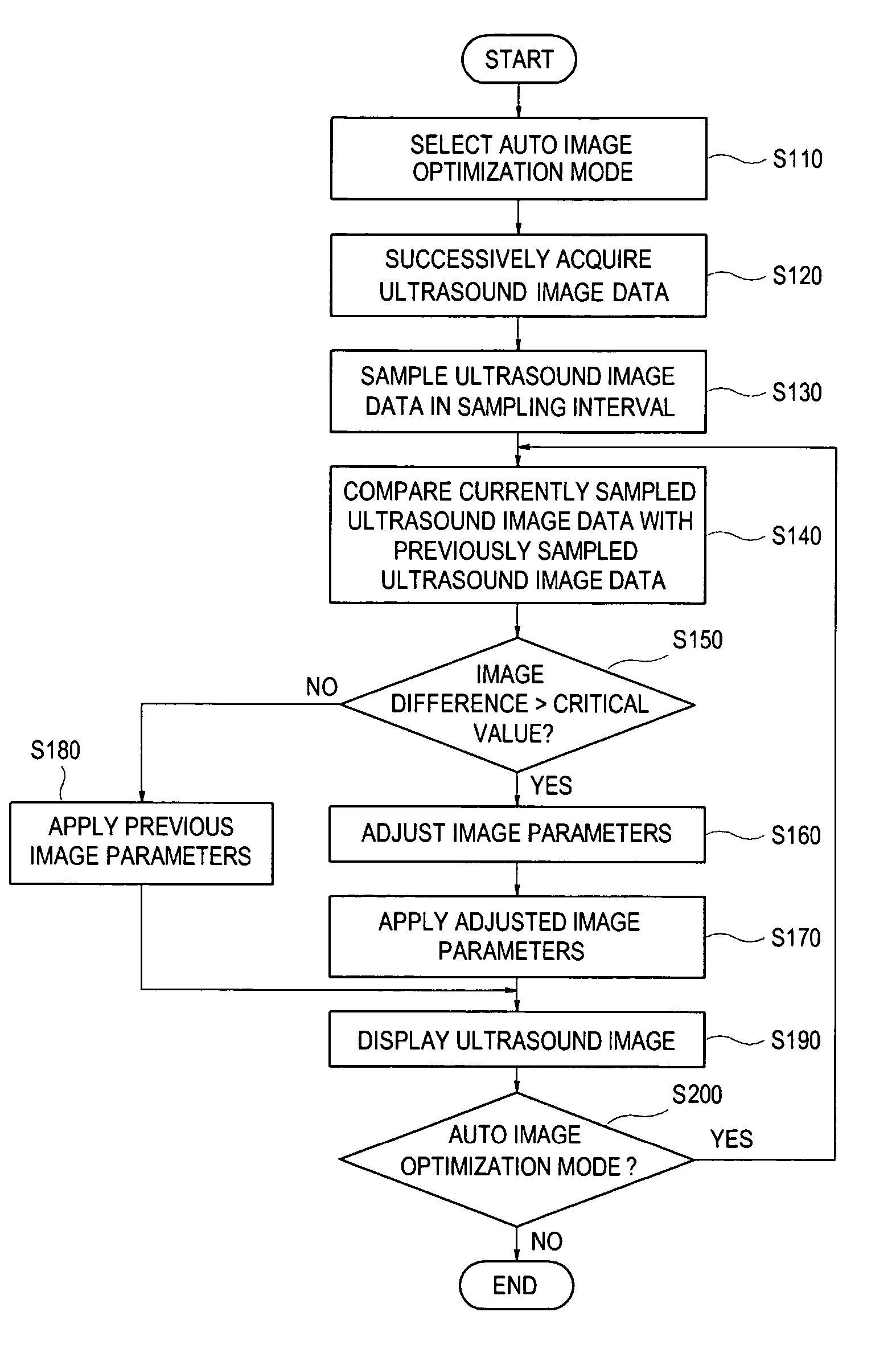
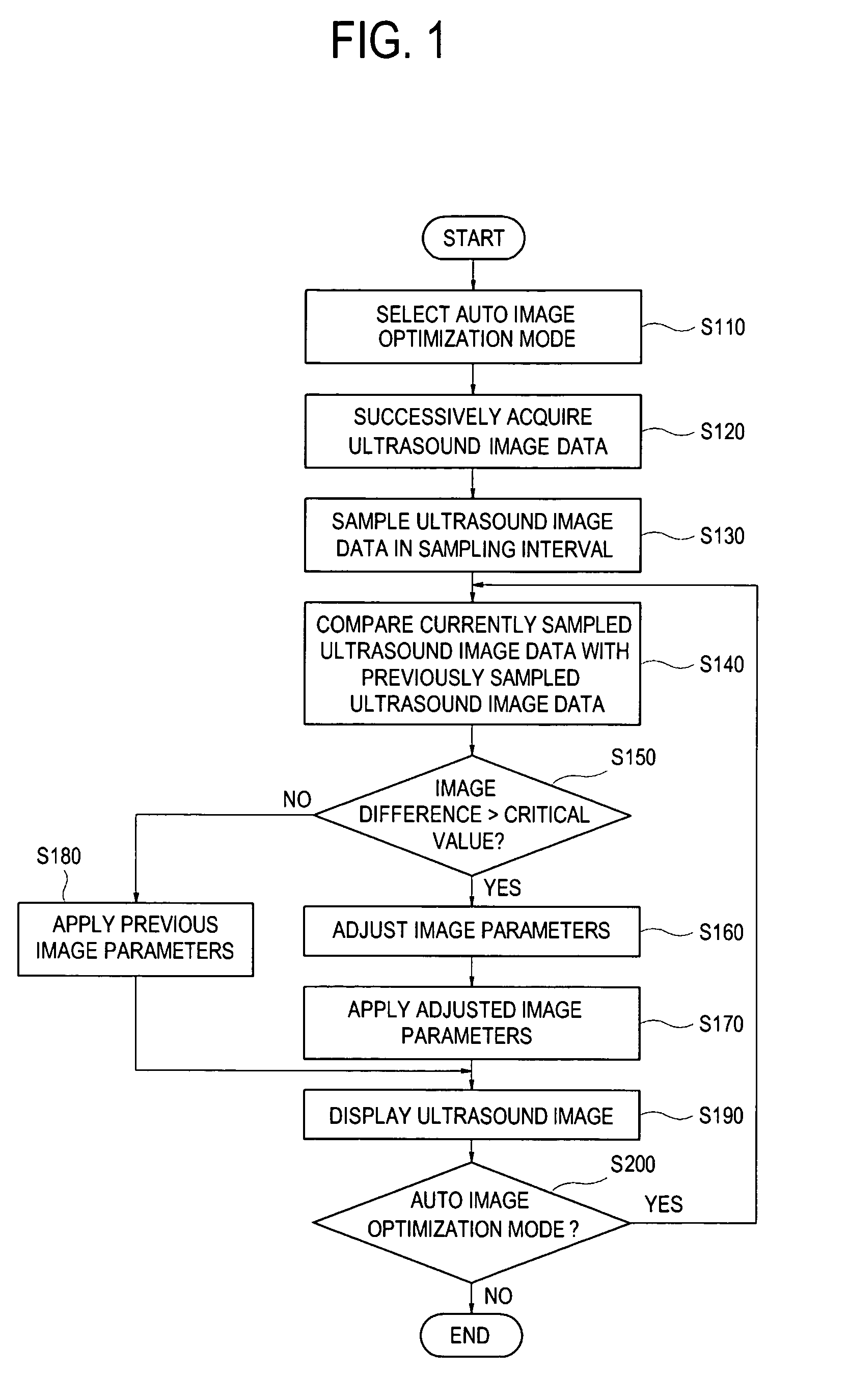
[0011]FIG. 1 is a flow chart showing a procedure for displaying an ultrasound image in accordance with a preferred embodiment of the present invention.

[0012] Referring to FIG. 1, if a sampling interval is set and then an auto image optimization mode is selected at step S110, then ultrasound image data are successively acquired from an object at step S120. The sampling interval represents a time interval to periodically sample an ultrasound image data from the successively acquired ultrasound image data. Further, the auto image optimization mode is a mode for automatically adjusting and optimizing image parameters such as gain, time gain, dynamic range (DR) to obtain an optimized ultrasound image. The sampling interval may be set within a range of 50 ms to 500 ms. The gain represents a value for adjusting amplification of a receive signal acquired from an ultrasound echo signal from the object. In addition, TG is a value for compensating a difference in strength of an ultrasound ech...


PUM


Abstract

The present invention is directed to a method of processing an ultrasound image, comprising the steps of: a) successively acquiring ultrasound images from an object; b) sampling ultrasound images in a predetermined time interval; c) determining an image difference between a currently sampled ultrasound image and a previously sampled ultrasound image; d) if the image difference is greater than a critical value, adjusting image parameters to optimize the image parameters; and e) applying the adjusted image parameters to the ultrasound images.

Description

FIELD OF THE INVENTION [0001] The present invention generally relates to a method of processing an ultrasound image, and more particularly to a method of automatically adjusting image parameters to optimize the same and applying the adjusted image parameters to the ultrasound image. BACKGROUND OF THE INVENTION [0002] An ultrasound diagnostic system has become an important and popular diagnostic tool since it has a wide range of applications. Specifically, due to its non-invasive and non-destructive nature, the ultrasound diagnostic system has been extensively used in the medical profession. Modem high-performance ultrasound diagnostic systems and techniques are commonly used to produce two or three-dimensional diagnostic images of internal features of an object (e.g., human organs). [0003] The ultrasound diagnostic system generally uses a wide bandwidth transducer to transmit and receive ultrasound signals. The ultrasound diagnostic system forms images of human internal tissues by e...

Claims


Application Information

Patent Timeline
Patent Type & Authority Applications(United States)
IPC IPC(8): G06K9/00
CPCG06T5/50A61B8/00
Inventor KIM, CHEOL ANAHN, CHI YOUNG
Owner MEDISON CO LTD
Learn More