A kind of biological tissue transparent imaging method

A biological tissue, transparent technology, applied in the field of biological sample processing and optical imaging, can solve the problems of unfavorable fluorescent protein retention, long operation time and high fat content in the transparentization time, so as to improve the transparentization efficiency, shorten the required time, and quickly transparent effect

Active Publication Date: 2020-08-18
HUAZHONG UNIV OF SCI & TECH
View PDF7 Cites 0 Cited by
  • Summary
  • Abstract
  • Description
  • Claims
  • Application Information

AI Technical Summary

Problems solved by technology

[0005] CUBIC technology achieves transparency through degreasing. The operation time of this technique is relatively long. Without staining the tissue, it takes at least 9 days to achieve transparency for a high-fat sample such as the liver. It seriously affects the efficiency of experimental data acquisition, and the longer the clearing time is, the more unfavorable it is to maintain the fluorescent protein, and the greater the possibility of losing sample imaging details (such as neuron axons, microglial antennae, etc.)
And CUBIC's refractive index matching reagent will have a certain impact on GFP fluorescence
Moreover, the liver, a special tissue sample, has a high fat content and a dense structure. It is still difficult for the existing clearing technology to achieve better clearing of the liver sample.

Method used


Image

  • A kind of biological tissue transparent imaging method
  • A kind of biological tissue transparent imaging method
  • A kind of biological tissue transparent imaging method

Examples


Embodiment 1

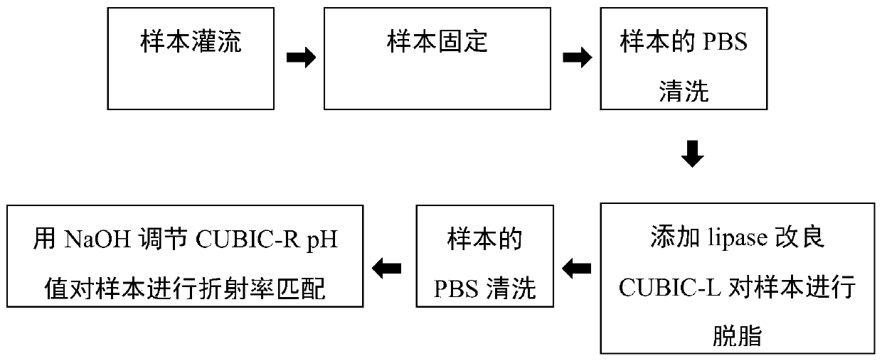
[0059] This embodiment provides a biological tissue transparent imaging method, which is an improvement on the CUBIC optical transparency scheme.

[0060] For the optical imaging application of CUBIC light-clearing technology in high-fat liver samples, we first optimized the protocol of CUBIC light-clearing technology. The sample used was the liver lobe of 8-week-old CX3CR1-mTmG mice. The steps included:

[0061] (1) Prepare 1×PBS: Prepare 1L 1×PBS with PBS powder, adjust the pH value to 7.4 with 1mol NaOH solution, then filter the prepared PBS with a filter membrane and put it at 4°C for later use.

[0062] (2) Preparation of 4% PFA: Prepare 4% PFA with prepared 1×PBS, adjust the pH value to 7.4, and store at 4°C.

[0063] (3) Prepare CUBIC-L reagent: 10% mass volume ratio of TritonX-100 and 10% mass volume ratio of N-butylethylenediamine, such as 40g TritonX-100 and 40g N-butylethylenediamine to constant volume Dissolve to 400mL ddH 2 O middle. Use a micrometer balance to...

Embodiment 2-4

[0074] Embodiments 2-4 respectively provide a biological tissue transparent imaging method, and the steps of each method are basically the same as those in Embodiment 1, and the differences are as follows:

[0075] In Example 2, the preparation method of CUBIC-L reagent A is: use a micrometer balance to weigh 100-500U / mg lipase powder and dissolve it into the prepared CUBIC-L, so that the lipase content reaches 4-20U / mL. All the other steps are consistent with Example 1.

[0076] In Example 3, after normal perfusion was performed on the dead mice, the PBS reagent containing saturated lipase was slowly perfused for 1 hour. The method of preparing lipase-containing PBS saturated reagent is as follows: add 0.4g 100-500U / mg lipase powder to 200mL 1×PBS, dissolve and mix well, filter with 200 / 400 mesh gauze to remove the insoluble part, and store at 4°C. Both CUBIC-L reagent A and CUBIC-L reagent B do not contain lipase, that is, the CUBIC-L reagent prepared in the same way as in ...

Embodiment 5

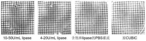
[0080] This example provides the optical imaging effect of the sample treated with the improved CUBIC scheme by adding lipase.

[0081] In order to detect the effect of the CUBIC protocol on fluorescence retention under different improved conditions, we performed confocal microscopic optical imaging on the liver lobes of CX3CR1-mTmG mice after clearing in Example 1 and Example 2:

[0082] We used the 10x water lens of LSM 780NLO confocal microscope (Zeiss, Germany) for imaging, and the excitation light was 488nm and 633nm. Under the same imaging conditions, different groups of samples were imaged fluorescently. Figure 5 It can be seen that each group is relatively good at maintaining RFP fluorescence, and the imaging depth can reach more than 2.6mm. However, in terms of the uniformity of RFP fluorescence retention of samples, the 4-20U / mL group has the best effect; for GFP As for the retention of fluorescence, it can be clearly seen that compared with the high-concentration l...


PUM


Abstract

The invention provides an improvement of a biological tissue transparence imaging technology and an application thereof. The method is an improvement of a CUBIC (Clear, unstructured Brain Imaging satellites and Computational Analysis) technology, and is mainly used to improve the transparence efficiency of the CUBIC light transparence technology and the retention of GFP fluorescence, and the method is named as f-CUBIC (fast fluorescent-CUBIC). The processing method comprises following steps that: pancreatic lipase is added into optically transparent reagent CUBIC-L to increase degreasing ability of CUBIC-L and improve transparentization efficiency, and in addition, NaOH is used for adjusting the pH value of the refractive index matching reagent CUBIC-R, so that the retention of the GFP fluorescence signal by the transparent tissue is improved. The reagent of the technology provided by the invention is easy to purchase, low in price and simple and convenient to operate, is particularlysuitable for processing high-fat-content tissues difficult to transparentize, such as livers, spleens and the like, is short in transparentization time and good in fluorescence maintaining effect, issuitable for polychromatic light transparentized fluorescence imaging, and is capable of improving the fluorescence imaging quality of samples, and enlarging the application range of the samples.

Description

technical field [0001] The invention belongs to the technical field of biological sample processing and optical imaging, and in particular relates to a biological tissue transparent imaging method. Background technique [0002] In recent years, the application of biological tissue optical transparency technology in biological imaging has become more and more popular, and has been widely used in large-volume imaging of intact tissues. For optical imaging of high-precision biological tissues, previous studies mainly focused on two-dimensional imaging of small areas of thin-layer samples, and the acquisition of three-dimensional imaging data mainly relied on layer-by-layer cutting imaging of samples. The development of biological tissue optical transparency technology combined with advanced optical imaging technology has played an important role in the three-dimensional imaging research of large tissue samples. It can transparentize opaque samples and reduce light scattering an...

Claims


Application Information

Patent Timeline
Patent Type & Authority Patents(China)
IPC IPC(8): G01N21/64
CPCG01N21/6428G01N21/6456G01N21/6458G01N21/6486
Inventor 张智红徐梦丽骆清铭
Owner HUAZHONG UNIV OF SCI & TECH
Why Eureka
Try Eureka