
A Method for Evaluating Fundus Image Quality

A fundus image, quality evaluation technology, applied in image analysis, image enhancement, image data processing and other directions, can solve the problems of fundus image evaluation, difficulty, lack of effective retinal information, etc., to achieve the effect of improving correlation and accurate automatic evaluation

Active Publication Date: 2021-08-03
北京云柿信息技术有限公司
View PDF10 Cites 0 Cited by
  • Summary
  • Abstract
  • Description
  • Claims
  • Application Information

AI Technical Summary

Problems solved by technology

[0003] Although the quality evaluation methods of fundus images are becoming more and more mature, most of them still rely on the subjective judgment of medical researchers. Although this subjective judgment method has certain reference value in evaluating the overall quality of fundus images , but it is difficult to evaluate the impact of fundus image quality on segmentation by subjective means
Fundus images can be used to extract blood vessel information, locate the optic disc, locate the macular area, etc. These applications provide huge information for the diagnosis and treatment of ophthalmic diseases, but the fundus images will inevitably be disturbed by noise during the acquisition and transmission process , leading to the loss of effective retinal information collected, which in turn affects the doctor's diagnosis

Method used


Image

  • A Method for Evaluating Fundus Image Quality

Examples


Embodiment Construction

[0039] The present invention will be further described in detail below in conjunction with the accompanying drawings and embodiments.

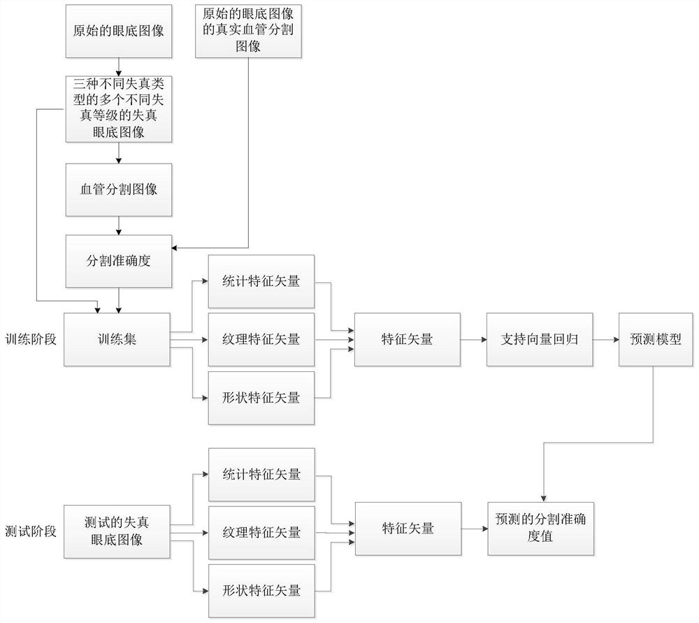
[0040] A fundus image quality evaluation method proposed by the present invention, its overall realization block diagram is as follows figure 1 As shown, it includes two processes of training phase and testing phase;

[0041] The specific steps of the described training phase process are:

[0042] ①_1. Select N original fundus images and the real blood vessel segmentation images of each original fundus image, and record the real blood vessel segmentation images of the uth original fundus image as M u ; Then each original fundus image is subjected to L different levels of fuzzy distortion, L different levels of overexposure distortion and L different levels of underexposure distortion, to obtain 3L pieces of distorted fundus corresponding to each original fundus image The images include L pieces of blurred and distorted fundus images, L piece...


PUM


Abstract

The invention discloses a fundus image quality evaluation method, which considers the influence of blurring, overexposure and underexposure on the segmentation accuracy of the fundus image, extracts statistical feature vectors, texture feature vectors, and shape feature vectors to form feature vectors, and then uses the support Vector regression trains the feature vectors of all distorted fundus images to construct a prediction model; in the test phase, by calculating the feature vectors of the distorted fundus images used for testing, and according to the prediction model constructed in the training phase, the distortion used for testing is predicted The segmentation accuracy value of the fundus image, because the obtained feature vector information can better reflect the change of the distortion of the distorted fundus image on the segmentation accuracy, so the predicted segmentation accuracy value and the real segmentation accuracy value are effectively improved. The correlation between the results, that is, the ability to accurately and automatically evaluate the quality of fundus images, effectively improves the correlation between objective evaluation results and subjective perception.

Description

technical field [0001] The invention relates to an image quality evaluation method, in particular to a fundus image quality evaluation method. Background technique [0002] The fundus image is captured by a special fundus camera. The fundus image includes the main physiological structures such as the optic disc, macula and blood vessels in the retina, and is an important type of image in medical imaging. Among them, the optic disc appears as a nearly circular bright color area in the normal fundus image, which has the strongest contrast with the background area, and is the initial area of ​​the optic nerve and blood vessels; the macula is rich in lutein, so it is in the normal fundus The image shows a dark area, and the dark area has no blood vessel structure, and there is an inward sunken area called the fovea in the center of the macula; the blood vessels start from the optic disc area and extend to the entire interior of the eyeball, presenting a tree-like distribution th...

Claims


Application Information

Patent Timeline
Patent Type & Authority Patents(China)
IPC IPC(8): G06T7/00G06T7/11
CPCG06T7/0012G06T2207/10004G06T2207/10024G06T2207/20081G06T2207/30041G06T2207/30168G06T7/11
Inventor 邵枫杨伟山李福翠
Owner 北京云柿信息技术有限公司
Learn More