
Locating expansion device of shoulder arthroscopy work passage

A technology of working channel and shoulder arthroscopy, which is applied in laparoscopy, medical science, surgery, etc., can solve the problems of affecting the progress of surgery, cumbersome operation, and waste of physical expenditure of doctors, so as to save physical labor expenditure, speed up the operation process, and meet the needs of patients. The effect of market demand

Active Publication Date: 2018-12-07
GENERAL HOSPITAL OF THE NORTHERN WAR ZONE OF THE CHINESE PEOPLES LIBERATION ARMY
View PDF6 Cites 3 Cited by
  • Summary
  • Abstract
  • Description
  • Claims
  • Application Information

AI Technical Summary

Problems solved by technology

[0002] Arthroscopic surgery is to insert a lens metal tube with a lighting device into the joint cavity through a small incision, and enlarge the internal structure of the joint cavity on a monitor to observe the pathological changes and locations in the joint cavity, and at the same time monitor the joint cavity under the monitor of the TV. Carry out a comprehensive inspection and clean up the lesion; the positioning and expansion of the arthroscope are generally carried out step by step. It is necessary to position the epidural puncture needle and then use the straight forceps to give the expansion channel, and then put the working cannula. The operation is cumbersome and affects the operation. Progress, during channel expansion, the doctor uses both hands to hold the straight forceps tightly, so that the straight forceps will stretch the channel. It is difficult for the doctor to control the force. Too little force will make it difficult to open the channel. Excessive force will easily cause the channel to expand too much, resulting in The surgical opening is too large to heal. At the same time, after the channel is expanded, the doctor still needs to hold the straight forceps firmly to maintain the opening state of the straight forceps and maintain the expanded state of the channel, which wastes a lot of physical expenditure of the doctor, increases the difficulty of the operation, and cannot meet the actual needs. We Invented a New Positioning Dilator for the Working Channel of Shoulder Arthroscopy

Method used


Image

  • Locating expansion device of shoulder arthroscopy work passage
  • Locating expansion device of shoulder arthroscopy work passage
  • Locating expansion device of shoulder arthroscopy work passage

Examples


Embodiment Construction

[0014] In order to make the purpose, technical solutions and advantages of the embodiments of the present invention clearer, the technical solutions in the embodiments of the present invention will be clearly and completely described below in conjunction with the drawings in the embodiments of the present invention. Obviously, the described embodiments It is a part of embodiments of the present invention, but not all embodiments. Based on the embodiments of the present invention, all other embodiments obtained by persons of ordinary skill in the art without creative efforts fall within the protection scope of the present invention.

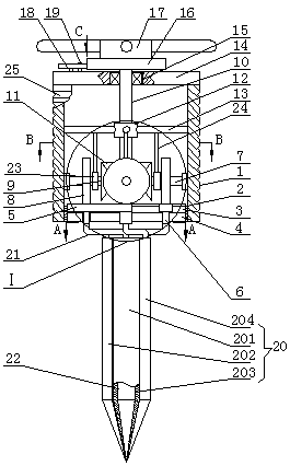
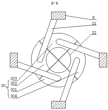
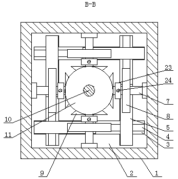
[0015]The shoulder arthroscopy working channel positioning dilator, as shown in the figure, includes a square tube 1, the bottom of the inner wall of the square tube 1 is fixedly installed with a lower plate 2, and the top surface of the lower plate 2 is provided with four strip chute 3, the strip chute 3 are connected in turn to form a well-shape...


PUM


Abstract

The invention discloses a locating expansion device of a shoulder arthroscopy work passage. The device comprises a square tube, a lower plate is fixedly installed at the bottom of the inner wall of the square tube, and four striped sliding grooves are formed in the top surface of the lower plate; the striped sliding grooves are successively connected to form a structureshaped like the Chinese character 'jing', striped through grooves are respectively formed in the bottom sides of the striped sliding grooves, and racks are respectively and movably installed in the striped sliding grooves and can respectively slide along corresponding striped through grooves; sliding blocks are respectively and movably installed in the striped through grooves and can respectively slide along corresponding striped through grooves, the top sides of the sliding blocks are respectively and fixedly connected to the ends of the bottom sides of the racks, and a cylinder is arranged right below a lower plate; the lower end of the cylinder body is in an inverted cone shape, the cylinder is composed of a front side pulling rod, a left side pulling rod, a back side pulling rod and a right side pulling rod, andthe shapes of the front side pulling rod, the left side pulling rod, the back side pulling rod and the right side pulling rod are same. The device is simple in structure, integrates an epiduralpuncture needle with straight pincers, after the epidural puncturing is completed, the expansion can be directly conducted on the passage, and the using and operation are convenient.

Description

technical field [0001] The invention belongs to the field of medical equipment, in particular to a positioning dilator for the working channel of shoulder arthroscopy. Background technique [0002] Arthroscopic surgery is to insert a lens metal tube with a lighting device into the joint cavity through a small incision, and enlarge the internal structure of the joint cavity on a monitor to observe the pathological changes and locations in the joint cavity, and at the same time monitor the joint cavity under the monitor of the TV. Carry out a comprehensive inspection and clean up the lesion; the positioning and expansion of the arthroscope are generally carried out step by step. It is necessary to position the epidural puncture needle and then use the straight forceps to give the expansion channel, and then put the working cannula. The operation is cumbersome and affects the operation. Progress, during channel expansion, the doctor uses both hands to hold the straight forceps ...

Claims


Application Information

Patent Timeline
Patent Type & Authority Applications(China)
IPC IPC(8): A61B17/00A61B1/317
CPCA61B1/317A61B17/00234A61B2017/0034
Inventor 刘欣伟刘松波巩天星周大鹏
Owner GENERAL HOSPITAL OF THE NORTHERN WAR ZONE OF THE CHINESE PEOPLES LIBERATION ARMY
Learn More