Space-time cooperation segmentation method based on infant brain tumor multi-modal MRI graph

A collaborative segmentation, infant technology, applied in the field of image processing and biomedicine, can solve the problems of difficult application of segmentation models and low tissue contrast, and achieve the effect of improving efficiency, accuracy and segmentation accuracy

Active Publication Date: 2017-04-26
WENZHOU MEDICAL UNIV
View PDF4 Cites 14 Cited by
  • Summary
  • Abstract
  • Description
  • Claims
  • Application Information

AI Technical Summary

Problems solved by technology

Therefore, it is difficult to apply the segmentation model of adult brain MR images to the segmentation of infant brain MR images
Especially at 6-8 months, the image contrast is in

Method used


Image

  • Space-time cooperation segmentation method based on infant brain tumor multi-modal MRI graph
  • Space-time cooperation segmentation method based on infant brain tumor multi-modal MRI graph
  • Space-time cooperation segmentation method based on infant brain tumor multi-modal MRI graph

Examples


Embodiment Construction

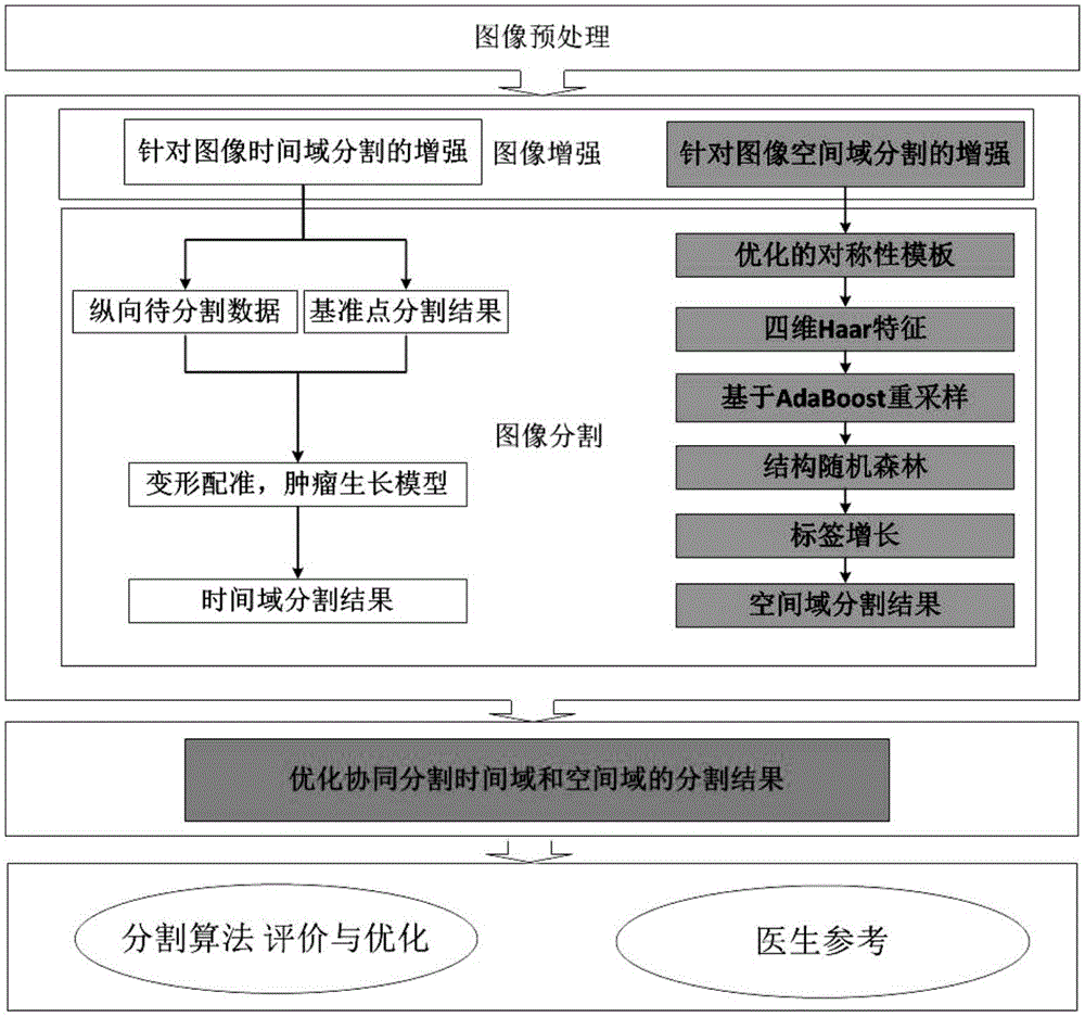
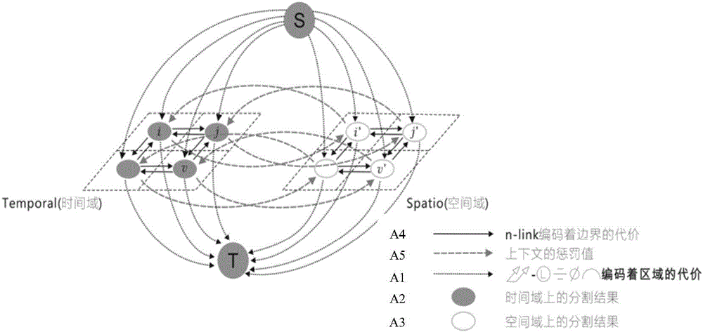
[0029] Specific embodiments of the present invention such as Figure 1-3 Shown is a space-time co-segmentation method based on multimodal brain tumor MRI image longitudinal data, which includes the following steps: (1) obtain the postoperative brain tumor MRI image, preprocess the image, (2) convert step (1) The longitudinal data of the method are respectively mapped to the time domain and the space domain for segmentation processing. (3) The time domain segmentation results and the space domain segmentation results are compared and referenced to construct a four-dimensional graph model.

[0030]Segmentation is performed according to the following steps: first, the entire tumor area is segmented, and the segmentation is performed according to steps (1)-(3); then, necrosis (Necrosis), enhancing tumor (Enhancing tumor), and non-tumor are segmented from the entire tumor area. Enhance the synthetic area of ​​the tumor (Non-enhancing tumor), exclude the edema (Edema) area, and foll...


PUM


Abstract

The invention discloses a space-time cooperation segmentation method based on an infant brain tumor multi-modal MRI graph. The space-time cooperation segmentation method includes (1) obtaining the postoperation brain tumor MRI images; (2) mapping the vertical data to the time domain and the spatial domain for segmentation; wherein the time domain segmentation includes obtaining the pre-operation segmentation result and the vertical data to be segmented, aligning the pre-operation and postoperation images, and constructing a postoperation tumor growth model; and the spatial domain segmentation includes constructing a healthy infant brain template, extracting the Haar structure characteristics, obtaining the preliminary probability result through the combination of a structure random forest method and an AdaBoost frame, increasing labels by means of similarity area increasing algorithm, and obtaining the spatial domain segmentation result; and (3) constructing a four-dimensional graph model through the combination of the time domain segmentation result and the spatial domain segmentation result, and optimizing the obtained parameters to form an automatic segmentation result. The segmentation method improves the accuracy of the infant brain tumor area segmentation.

Description

technical field [0001] The invention belongs to the technical field of combining image processing and biomedicine, in particular to a space-time collaborative segmentation method based on multimodal MRI images of infantile brain tumors. Background technique [0002] Brain tumors refer to cancerous substances growing in the cranial cavity, including primary tumors caused by lesions in the brain parenchyma, and secondary tumors that metastasize and invade the brain from other parts of the body. Segmentation is an important prerequisite for brain tumor diagnosis, surgical planning, radiotherapy and chemotherapy, and long-term longitudinal studies. Among various medical imaging techniques, magnetic resonance imaging (MRI) has a strong resolution of soft tissues, so it is more accurate to define tumor boundaries based on this. Brain tumor segmentation in MRI images is of great significance in diagnosis, pathological analysis, treatment and scientific research. Clinically, exper...

Claims


Application Information

Patent Timeline
IPC IPC(8): G06T7/149G06T7/11G06T7/194
CPCG06T2207/10088G06T2207/30016
Inventor 潘志方叶夏王贤川应一凡陈峰
Owner WENZHOU MEDICAL UNIV
Why Eureka
Try Eureka