
Method for positioning three axial positions of fetal brain through nuclear magnetic resonance

A magnetic resonance and fetal technology, applied in the field of medical image processing and positioning

Inactive Publication Date: 2013-06-05
THE FIRST AFFILIATED HOSPITAL OF THIRD MILITARY MEDICAL UNIVERSITY OF PLA
View PDF4 Cites 0 Cited by
  • Summary
  • Abstract
  • Description
  • Claims
  • Application Information

AI Technical Summary

Problems solved by technology

[0003]Analyze the main reason, because the position of the fetus in the mother’s body is ever-changing, the correct position of the fetus cannot be directly obtained from the mother’s body, which eventually leads to difficulties in scanning and positioning

Method used


Image

  • Method for positioning three axial positions of fetal brain through nuclear magnetic resonance
  • Method for positioning three axial positions of fetal brain through nuclear magnetic resonance
  • Method for positioning three axial positions of fetal brain through nuclear magnetic resonance

Examples


Embodiment Construction

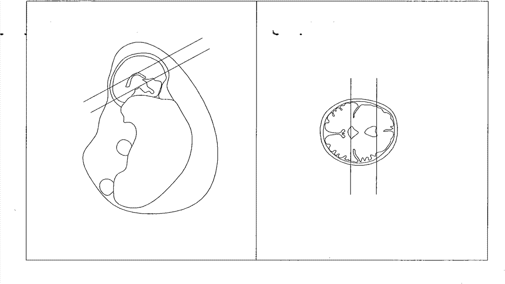
[0026] The first step is to locate the coronal and sagittal positions of the mother's uterus on the scout image;

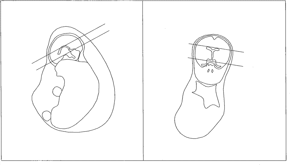
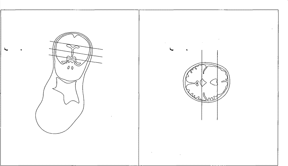
[0027] The second step is to obtain the preliminary body position image of the fetus from the image of the first step; due to reasons such as positioning lines and fetal positions, the positioning image of the second step shows that the shape and position of the fetal head are different, that is, the following two types may occur: Condition:

[0028] In the first case, when there is a standard fetal triaxial position, the midline of the fetal skull, the line connecting the lower edge of the genu of the corpus callosum and the lower edge of the splenium, and the line connecting the bottom of the bilateral temporal lobes can be observed, such as figure 1 As shown, if the fetal sagittal and axial positions appear during the scan, the standard coronal position of the fetus should be positioned first, which is parallel to the brainstem in the sagittal position and p...


PUM


Abstract

The invention relates to a method for positioning three axial positions of a fetal brain through magnetic resonance. The method is used for further rapidly, accurately and normatively positioning an image of the three axial positions of the fetal brain on the basis of the traditional nuclear magnetic resonance scanning and positioning technology and providing a more accurate and normative cross-section image for clinical medicine. Because the position and the direction of a fetus in a parent are possibly changed at any time, the standard three axial positions of only a few fetuses can be directly positioned through the standard three axial positions of the parent; in many cases, the position of the fetus in the parent is changeable; the standard image of the three axial positions cannot be obtained through the traditional technology; when the standard three axial positions of the parent cannot be given or the standard three axial positions of the fetus cannot be better given, a series of images of various positions can be obtained by rotating and positioning in 360 DEG through a haste sequence, namely a heavy T2 weighted line; and the image closest to the standard fetal position is selected from the series of images and served as the standard to position the brain.

Description

technical field [0001] The invention relates to a medical image processing and positioning method, in particular to a method for positioning the three-axis position of the fetal brain based on fetal nuclear magnetic resonance imaging scanning. [0002] Background technique [0003] So far, due to the different positions of the fetus in the mother's body, the standard triaxial position of the fetus and the standard triaxial position of the mother are different. It is impossible to accurately locate the standard triaxial position of the fetus on the standard triaxial position of the mother by using nuclear magnetic resonance. axial position. Therefore, it is often impossible to obtain the most satisfactory images when relying on the standard triaxial images of the mother to reconstruct the triaxial images of the fetal brain. Under normal circumstances, image processing is mostly based on the clinical experience of doctors. Due to factors such as human error, the final three-a...

Claims


Application Information

Patent Timeline
Patent Type & Authority Patents(China)
IPC IPC(8): A61B5/055A61B19/00A61B90/14
Inventor 蔡萍毛天骐赖力王新王健
Owner THE FIRST AFFILIATED HOSPITAL OF THIRD MILITARY MEDICAL UNIVERSITY OF PLA
Learn More