
Proctoscopic ultrasonic Doppler probe

A technology of rectoscopy and Doppler, applied in the direction of catheter, application, internal bone synthesis, etc., can solve the problem of single function, and achieve the effect of effective and convenient treatment of hemorrhoids

Inactive Publication Date: 2010-09-15
南京奥珂森电子有限公司
View PDF3 Cites 0 Cited by
  • Summary
  • Abstract
  • Description
  • Claims
  • Application Information

AI Technical Summary

Problems solved by technology

Therefore, the function of the original proctoscope ultrasonic Doppler probe has shown certain limitations, and it is no longer suitable for suturing and suspending hemorrhoids. There is a problem of single function, which cannot meet the requirements of modern treatment technology.

Method used


Image

  • Proctoscopic ultrasonic Doppler probe
  • Proctoscopic ultrasonic Doppler probe
  • Proctoscopic ultrasonic Doppler probe

Examples


Embodiment 1

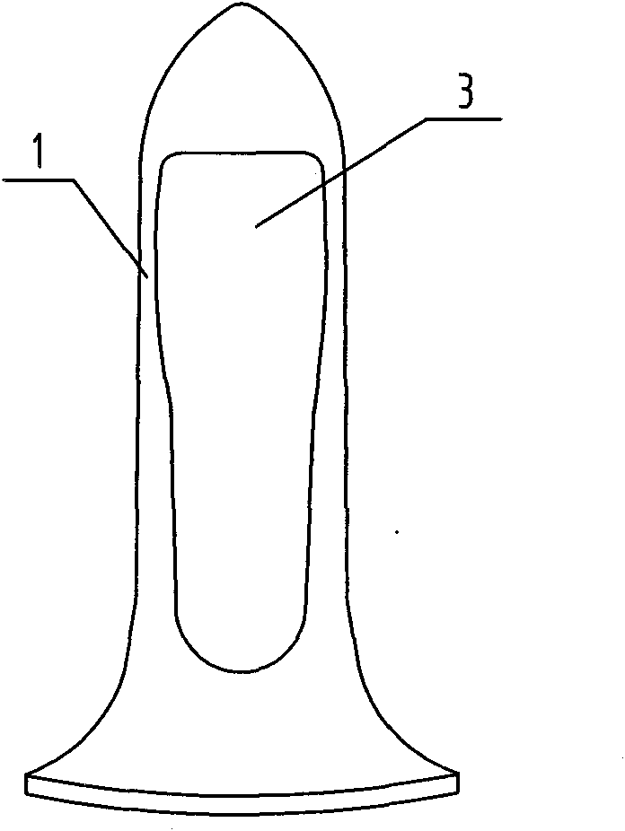
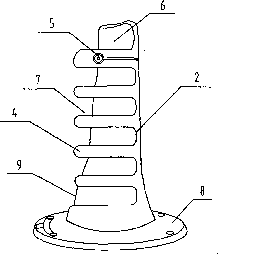
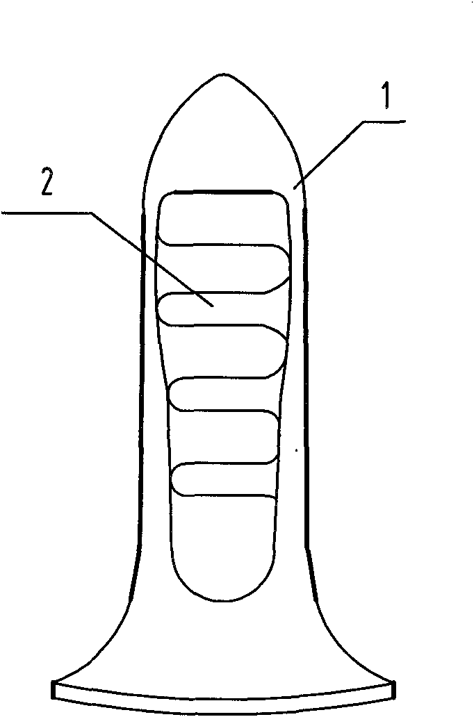
[0019] This embodiment is an ultrasonic Doppler probe for a rectoscope, which is of a split structure and consists of an outer casing 1 and a probe body 2 matching the inner space of the outer casing. The structure of the outer casing 1 is as figure 1 As shown, its profile is bullet-shaped, and a treatment window 3 is arranged on the outer casing 1, and the treatment window 3 is a tooth shape with a slightly larger upper part and a slightly smaller lower part. The structure of the probe body 2 is as figure 2 As shown, a group of fence-shaped bifurcation bodies 4 extending in the circumferential direction are arranged at intervals on the probe body 2, wherein an ultrasonic transducer 5 is installed on a bifurcation body adjacent to the end of the outer casing 1, and is connected to the treatment window. The hemorrhoidal artery ligation operation area 6 is formed between the edges of 3, and the hemorrhoid body suture suspension operation area 7 is formed between the intervals ...

Embodiment 2

[0024] The structure of this embodiment is similar to that of Embodiment 1, the difference lies in the relative fixation of the outer shell 1 and the probe body 2, and the probe body 2 of this embodiment is also supported by an arc extending axially from the flange mouth 8. 9 and the fence-shaped bifurcation body 4 extending from the arc brace along the circumferential direction, the envelope shape of the arc brace 9 and the bifurcation body 4 has a tapered taper, and each bifurcation body 4 is located in the same outer cone on the helix of the thread, such as Figure 5 As shown, the inner wall of the outer casing 1 has a matching internal tapered thread. In this way, when the probe body 2 is set in the outer shell 1, the fence-shaped branch body 4 is rotated to the treatment window 3, and then rotated by a small angle, the thread of the branch body 4 can be threaded with the inner taper thread of the inner wall of the outer shell 1. occluded to achieve the relative fixation ...


PUM


Abstract

The invention relates to a clinical medical probe, in particular to a proctoscopic ultrasonic Doppler probe which has a splitting-type structure and comprises a bullet-shaped outer shell and a probe body which is matched with the internal space of the outer shell, wherein a treatment window is arranged on the outer shell, a plurality of annular forking bodies with certain intervals are evenly distributed on the probe body, an ultrasonic energy transducer is arranged on one forking body at the upmost end, a hemorrhoidal artery perstriction area is arranged above one forking body positioned at the upmost end, and hemorrhoid suture suspension areas are formed at interval spaces among all forking bodies. As the plurality of annular forking bodies with certain intervals are scientifically arranged on the probe body, the hemorrhoidal artery perstriction area and a plurality of hemorrhoid suture suspension areas are formed in a very reasonable layout, therefore, the joint of hemorrhoidal artery perstriction and hemorrhoid suture suspension which are carried out on the same probe is smartly solved, and the hemorrhoid treatment is more effective and convenient.

Description

technical field [0001] The invention belongs to the technical field of medical instruments, and relates to a clinical medical probe, in particular to a proctoscope ultrasonic Doppler probe. Background technique [0002] The previous proctoscope ultrasonic Doppler probe was a single bullet-shaped probe body, which was provided with a treatment window and an ultrasonic transducer installed on the probe body. When treating hemorrhoids, the transducer was used to transmit ultrasonic waves and receive ultrasonic waves to The location of the hemorrhoidal artery is detected, and then the hemorrhoidal artery is ligated at the treatment window to treat hemorrhoids. [0003] With the advancement of medical technology and the improvement of surgical requirements, in the latest hemorrhoid treatment technology, not only to find the hemorrhoid arteries, ligate the hemorrhoid arteries, but also suture and suspend the hemorrhoids. Therefore, the function of the original proctoscope ultraso...

Claims


Application Information

Patent Timeline
Patent Type & Authority Applications(China)
IPC IPC(8): A61B8/12A61B17/94
Inventor 钱旭波陈学军
Owner 南京奥珂森电子有限公司
Learn More