Method for detecting disease-related marker using gastric mucosal lavage fluid
A technology of markers and gastric mucosa, which is applied in the field of detection of disease-related markers, can solve problems such as retention, inability to extract Southern blots, diagnostic possibility and diagnostic accuracy, and achieve the effect of simple collection and avoiding pollution
- Summary
- Abstract
- Description
- Claims
- Application Information
AI Technical Summary
Problems solved by technology
Method used
Image

Examples
Embodiment 1
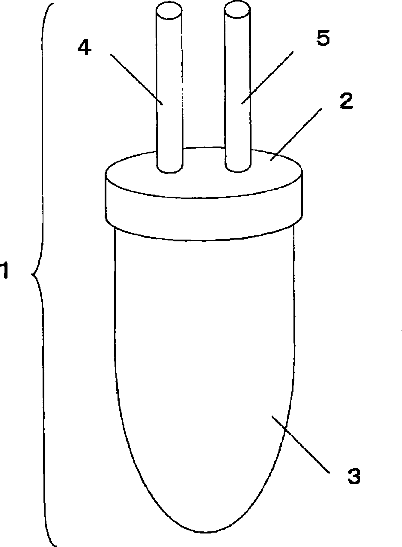
[0088] Embodiment 1: Specimen collection container
[0089] figure 1 An example of the sample collection container of the present invention is shown. The sample collection container 1 includes a cap-shaped sample collection container upper part 2 and a sample collection container lower part 3 with a capacity of 50 ml, both of which are airtightly joined by screwing. Two connecting parts, ie, a scope-side connecting part 4 and an aspirator-side connecting part 5, are provided on the upper part 2 of the sample collection container, and can be airtightly connected to a suction duct of an endoscope apparatus. In addition, 0.5 ml of 0.5 M EDTA (Wako Pure Chemical Industries, Ltd. K.K. special grade product) was sealed in the sample collection container 1 .
Embodiment 2
[0090] Example 2: Preparation of Specimen
[0091] Remove the suction tube connected to the connection part from the suction bottle of the endoscope device (EVIS LUCERA, Olympus Corporation), connect it to the scope-side connection part of the above-mentioned sample collection container, and simultaneously collect the sample The suction side connection part of the container is connected with the scope side connection part of the suction bottle to form a closed circuit.
[0092] Dilute 4 ml of simethicone (Gascon (registered trademark) drops, Kissei Pharmaceutical Co., Ltd.) (containing 80 mg of dimethyl polysiloxane) with 50 to 100 ml of ordinary water, and dissolve 20,000 units in the diluted solution Pronase (registered trademark) (Pronase (registered trademark) MS, Kakuken Pharmaceutical Co., Ltd.) and 1 g of baking soda were administered to human subjects suffering from various stomach problems, and after 10 to 15 minutes, the subjects were placed in a supine position. ...
Embodiment 3
[0095] Example 3: Evaluation of DNA Quality and Quantity
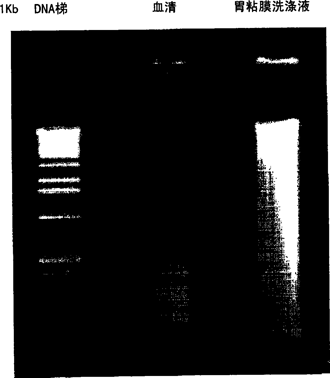
[0096] i) Evaluation of DNA quality
[0097] The quality of DNA contained in the above samples derived from human subjects with gastric cancer was evaluated by electrophoresis. In the electrophoresis method, 1-2 μg of DNA sample was dissolved in 10 μl of H 2 Diluted in O, run electrophoresis on 1% agarose gel at 100V for 30 minutes. The results of electrophoresis evaluation were as follows: figure 2 shown. In the figure, the left lane is a positive control (1Kb DNA Extension Ladder, Cat. No. 10511-012, Invitrogen), the middle lane is a serum sample, and the right lane is a gastric mucosa washing solution. Either result showed that gastric mucosal wash samples contained DNA of better quality than serum samples and comparable to biopsy samples.
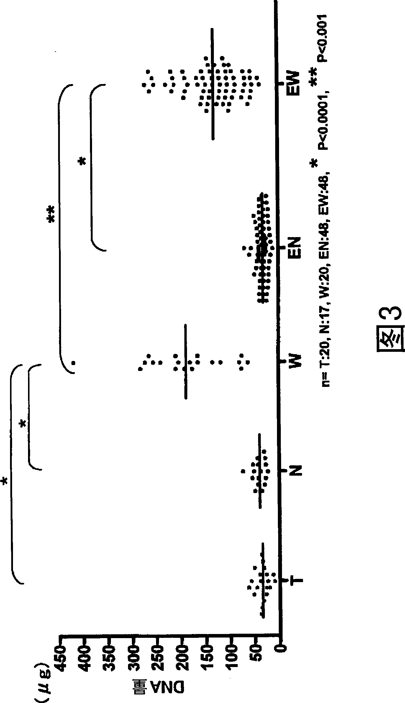
[0098] ii) Evaluation of DNA amount
[0099] The recovered amount of DNA contained in samples derived from human subjects with gastric cancer was evaluated spectrophoto...
PUM
 Abstract
Description
Claims
Application Information
 - R&D
- Intellectual Property
- Life Sciences
- Materials
- Tech Scout
- Unparalleled Data Quality
- Higher Quality Content
- 60% Fewer Hallucinations
Browse by: Latest US Patents, China's latest patents, Technical Efficacy Thesaurus, Application Domain, Technology Topic, Popular Technical Reports.
© 2025 PatSnap. All rights reserved.Legal|Privacy policy|Modern Slavery Act Transparency Statement|Sitemap|About US| Contact US: help@patsnap.com