Combined segmentation and registration framework for parametric shapes

a parametric shape and registration framework technology, applied in image analysis, image enhancement, instruments, etc., can solve the problems of no known method or apparatus to model lesion segmentation and correspondence simultaneously, and the difficulty of detecting and segmenting those lesions, so as to maximize the probability of p

Active Publication Date: 2011-11-22
SIEMENS HEALTHCARE GMBH
View PDF1 Cites 5 Cited by
  • Summary
  • Abstract
  • Description
  • Claims
  • Application Information

AI Technical Summary

Benefits of technology

[0012]As yet another aspect of the present invention, a method is provided, further comprising marking each object i in image I, generating and optimizing a segmentation contour for each object i of N objects in baseline image I, the contour of each object in I being determined by one or more contour parameters γi, generating a contour for each object i′ of N objects in image I′, the contour of each object I′ being determined by one or more contour parameters γi′, optimizing segmentation and registration of the i′ of N objects in image I′ by maximizing the conditional probability expression P(Γ′|Γ,I′)∝P(I′|Γ,Γ′)P(Γ|Γ′)P(Γ′), wherein Γ=(γ1, . . . , γN) represents the one or more contour parameters of each of the N objects in the image I, and Γ′=(γ′1, . . . , γ′N) represents the one or more parameters of each of the N objects in image I′, and generating an image for display with segmented and registered objects identified.
[0018]As yet another aspect of the present invention a method is provided, wherein generating the segmentation contour for each object i of N objects in baseline image I, comprises providing a click-point x0i to each object i of the N objects in image I to create N click-points X0=(x01, . . . ,x0N), assigning a prior shape to each contour, parameterizing a level-set function φγ representing a shape, based on the prior shape, and maximizing a conditional probability P(Γ|I,X0).

Problems solved by technology

As each contrast phase varies dramatically in the amount of contrast agent that is visible in the image, the ability to detect and segment those lesions becomes extremely challenging in some phases for a segmentation application operating on a computer and even for the human eye.
One of the challenges is heavily varying contrast between the phases.
Another challenge is the apparent deformations of objects between the follow-up studies.
It is believed that currently there are no known methods or apparatus to model lesion segmentation and correspondence simultaneously in a combined framework.

Method used


Image

  • Combined segmentation and registration framework for parametric shapes
  • Combined segmentation and registration framework for parametric shapes
  • Combined segmentation and registration framework for parametric shapes

Examples


Embodiment Construction

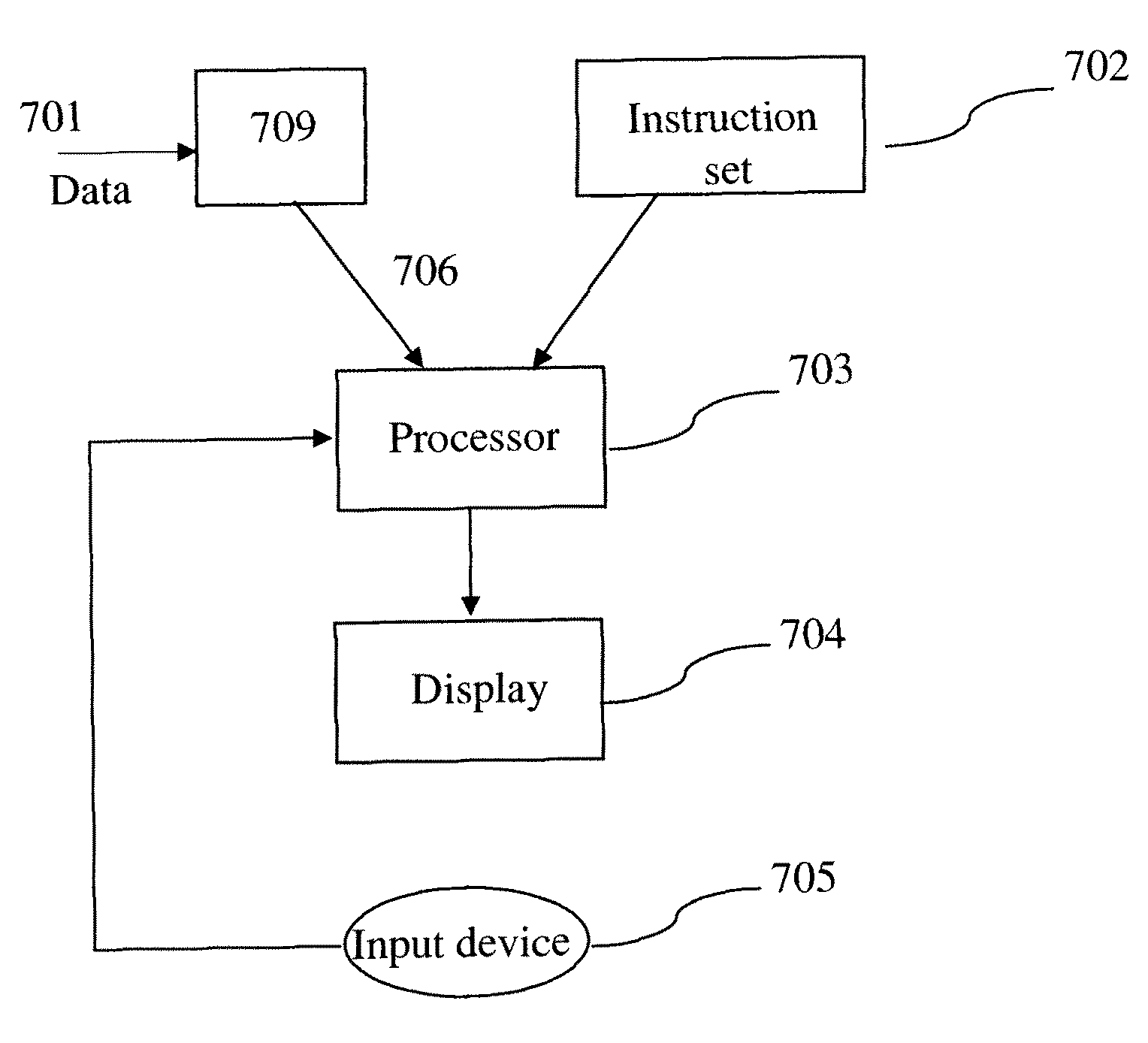
[0030]Novel methods and apparatus for segmentation and registration of general shapes and objects and as applied to applications such as medical images are provided herein. The application of CT multi-phase follow-up studies is used to describe an embodiment of the general approach. Aspects of the invention are intended to cover both the model description and its application to oncology care cancer monitoring, such as in an imaging system.

[0031]The provided framework is composed in one part of a novel region based parametric level-set segmentation formulation.

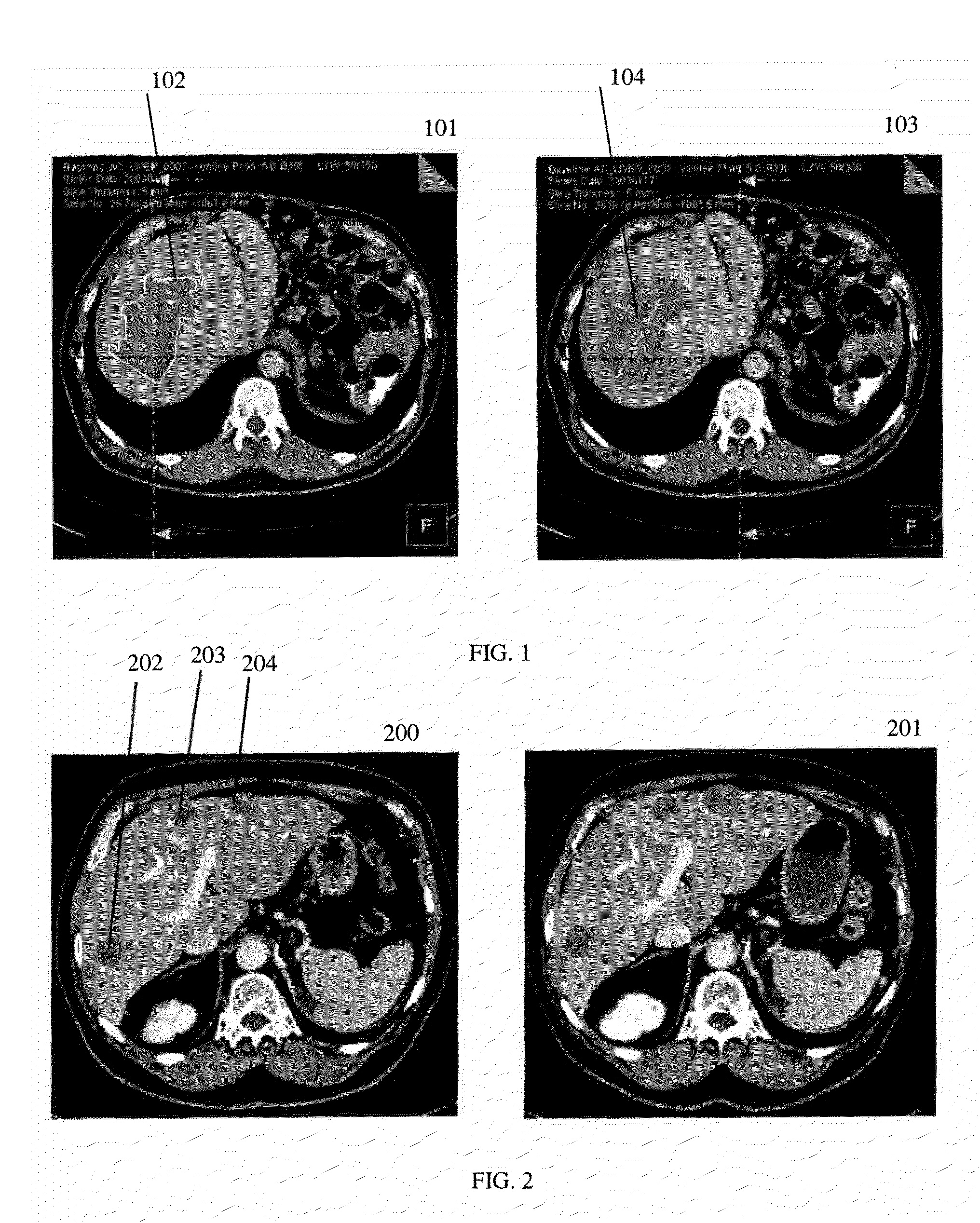
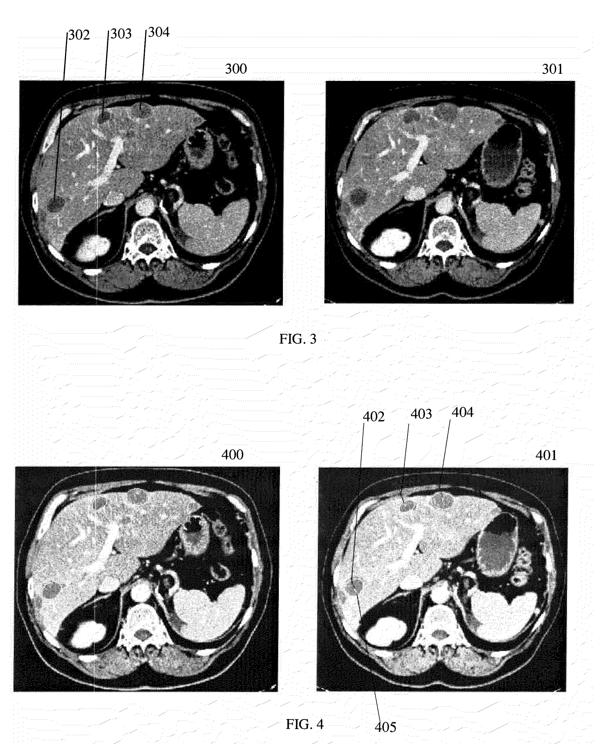
[0032]In one embodiment of the present invention the problem of locating and measuring previously identified lesions in one or more follow-up images is solved. An exact registration of the datasets is not necessary; this is implicitly done by a method that will be provided next. A possible workflow could be as follows.

[0033]1. Manual initialization—Looking at one or several images of the baseline study, a user such as a physici...


PUM


Abstract

Progress monitoring of lesions is done automatically by segmentation and registration of lesions in multi-phase medical images. A parametric level-set framework includes a model optimization for any number of lesions. The user specifies lesions in a baseline volume by clicking inside of them. The apparatus segments the lesions automatically in the baseline and follow-up volumes. The segmentation optimization compensates for lesion motion between baseline and follow-up volumes. 2D and 3D medical patient data can be processed by the methods.

Description

CROSS-REFERENCE TO RELATED APPLICATIONS[0001]This application claims the benefit of U.S. Provisional Application No. 61 / 105,498, filed Oct. 15, 2008, which is incorporated herein by reference.BACKGROUND OF THE INVENTION[0002]The present invention relates to methods and apparatus for image segmentation and image registration. In particular, it relates to segmentation and registration of medical 2D and 3D images that are acquired at different phases and / or at different times. Imaging applications for oncology follow-up studies have just recently been introduced to the clinical workflow. Cancer treatment has also become an extraordinary important procedure in today's healthcare due to cancer being among the most frequent causes of death. Monitoring the patient's response to radiation treatment by investigating cancer development is crucial for determining success or failure of the therapy. One indicator for cancer development is the change in size and structure of lesions found in thor...

Claims


Application Information

Patent Timeline
Patent Type & Authority Patents(United States)
IPC IPC(8): G06K9/00
CPCG06K9/342G06K9/6207G06T7/0032G06T7/0034G06T7/0081G06T7/0097G06T2207/10081G06T2207/20101G06T2207/20141G06T2207/20161G06T2207/30056G06T7/344G06T7/35G06T7/11G06T7/174G06T7/187G06V10/267G06V10/755
Inventor KIRCHBERG, KLAUS J.GUETTER, CHRISTOPH
Owner SIEMENS HEALTHCARE GMBH
Why Eureka
Try Eureka