
Systems, methods and computer-accessible medium which provide microscopic images of at least one anatomical structure at a particular resolution

a microscopic image and resolution technology, applied in the field of imaging systems, apparatuses and methods, can solve the problems of difficult or impossible interrogation, affecting the diagnosis of cad, and unable to achieve the resolution of images in only a narrow depth rang

Active Publication Date: 2011-10-27
THE GENERAL HOSPITAL CORP
View PDF11 Cites 27 Cited by
  • Summary
  • Abstract
  • Description
  • Claims
  • Application Information

AI Technical Summary

Benefits of technology

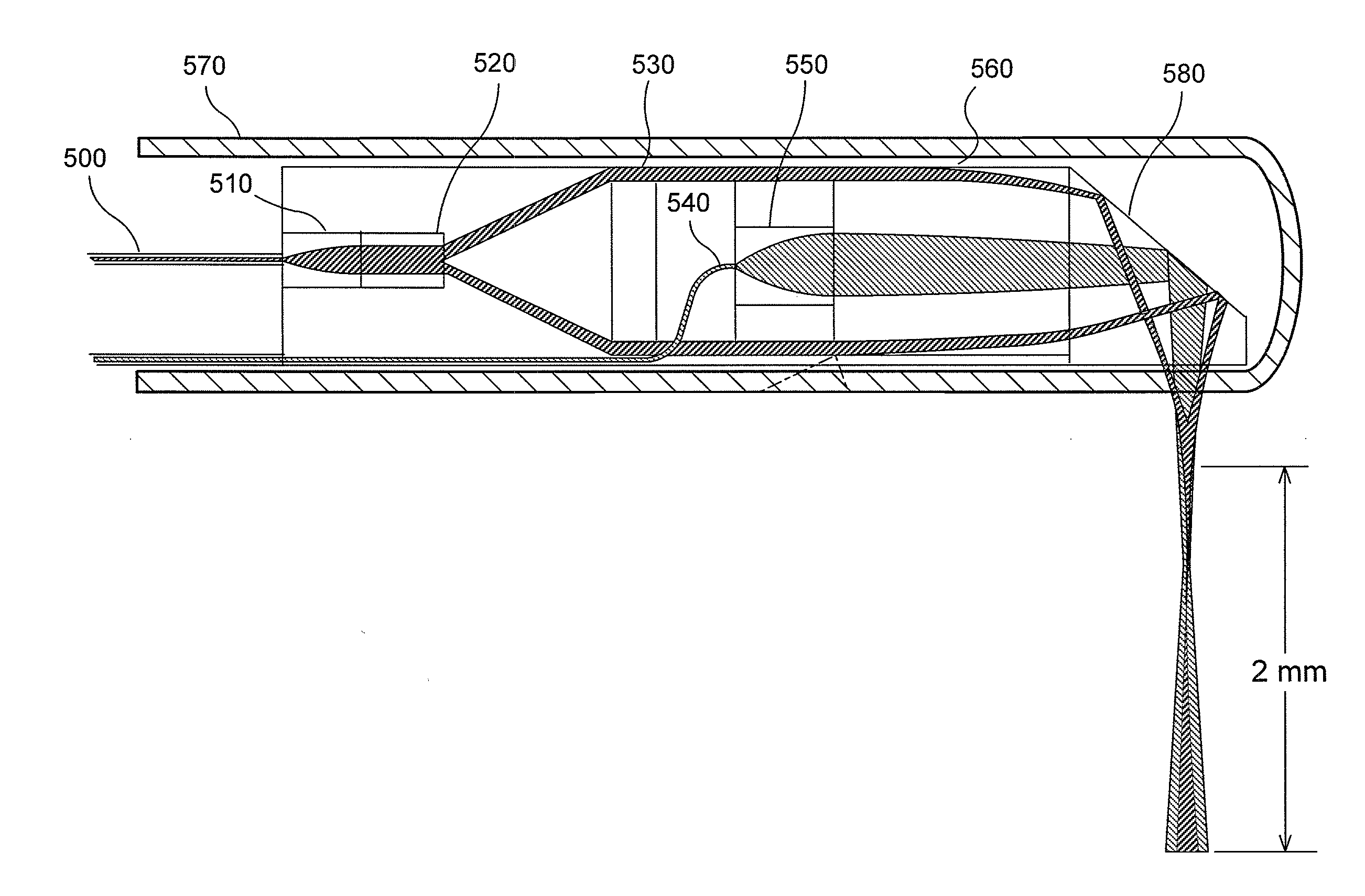
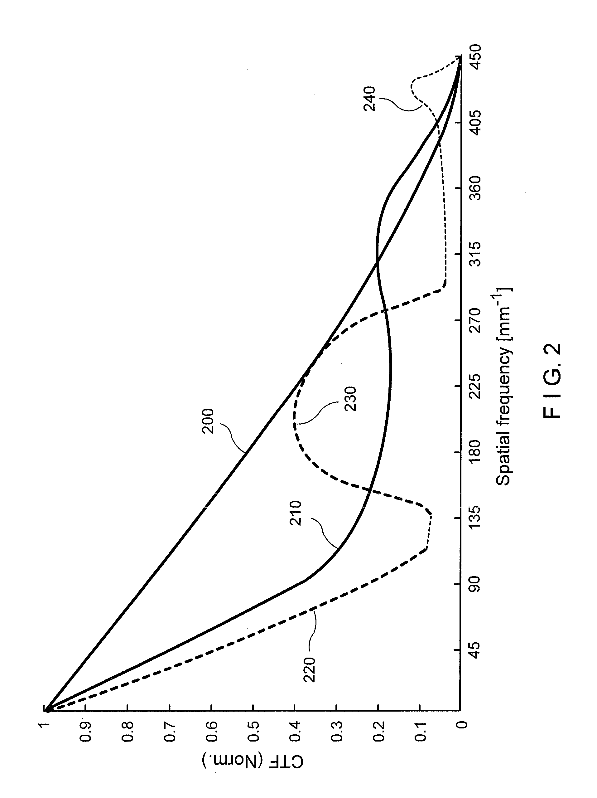
[0011]According to another exemplary embodiment of the present disclosure, more than two imaging channels can illuminate / detect different Bessel and / or Gaussian beams. In yet a further exemplary embodiment, different transfer functions can be illuminated and / or detected. The exemplary combination of images obtained with such additional exemplary beams can facilitate the μOCT CTF to be provided to the diffraction-limited case, and can also facilitate a depth-of-field extension even further.

Problems solved by technology

Certain progress in understanding, diagnosing, and treating CAD has been hindered by the fact that it has been difficult or impossible to interrogate the human coronary wall at cellular-level resolution in vivo.
This conventional method, however, neglects the intrinsic compromise between transverse resolution and depth of field in cross-sectional OCT images and results in images in which only a narrow depth range is resolved.
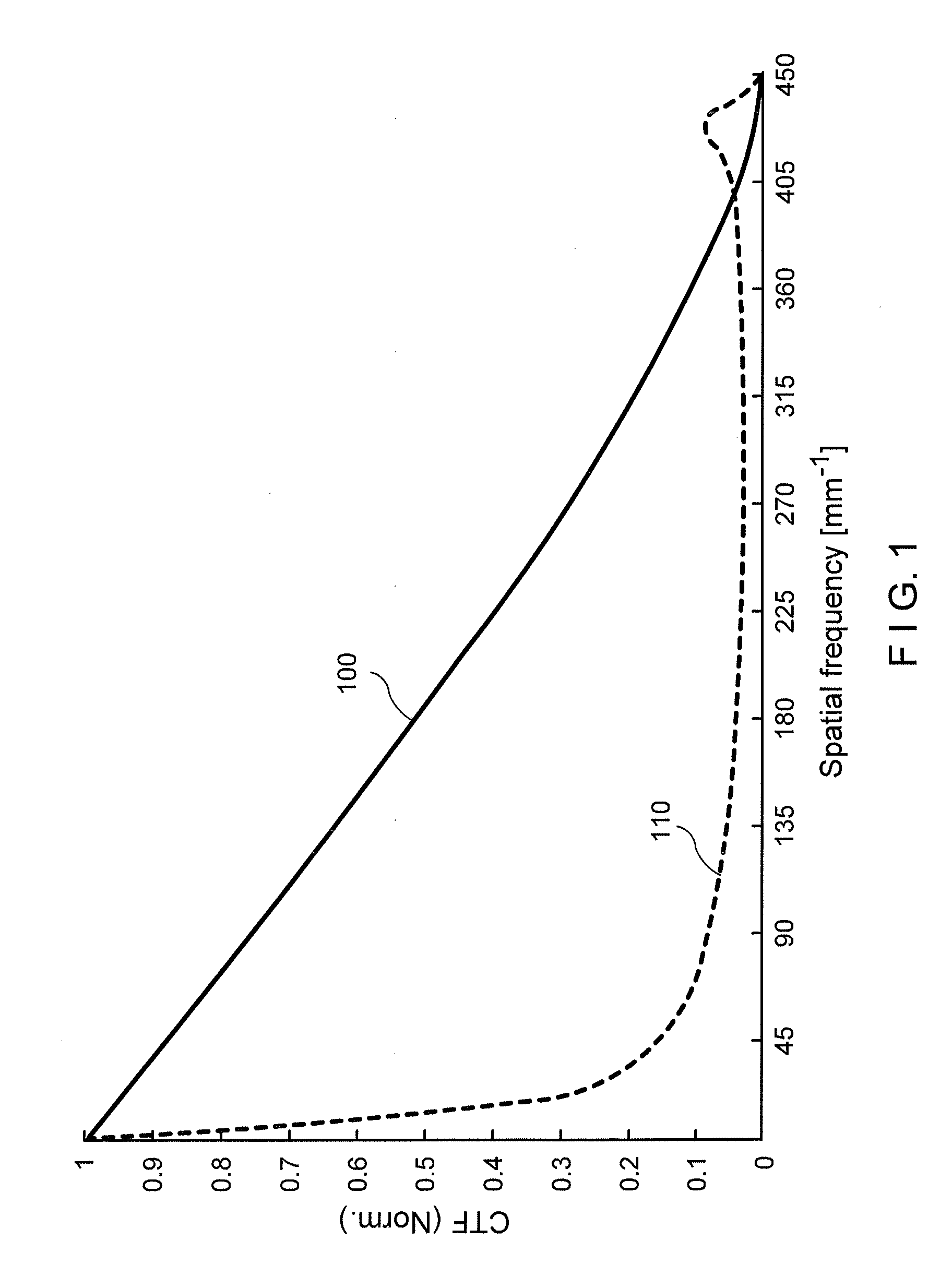
Bessel beam illumination and detection of light reflected from the sample, however, can suffer from a significant reduction in contrast and detection efficiency.

Method used


Image

  • Systems, methods and computer-accessible medium which provide microscopic images of at least one anatomical structure at a particular resolution
  • Systems, methods and computer-accessible medium which provide microscopic images of at least one anatomical structure at a particular resolution
  • Systems, methods and computer-accessible medium which provide microscopic images of at least one anatomical structure at a particular resolution

Examples


Embodiment Construction

[0010]To address and / or overcome such deficiencies, one of the objects of the present disclosure is to provide exemplary embodiments of systems, methods and computer-accessible medium according to the present disclosure, which can provide microscopic images of at least one anatomical structure at a particular resolution. Another object of the present disclosure is to overcome a limited depth of focus limitations of conventional Gaussian beam and spatial frequency loss of Bessel beam systems for OCT procedures and / or systems and other forms of extended focal depth imaging.

[0011]According to another exemplary embodiment of the present disclosure, more than two imaging channels can illuminate / detect different Bessel and / or Gaussian beams. In yet a further exemplary embodiment, different transfer functions can be illuminated and / or detected. The exemplary combination of images obtained with such additional exemplary beams can facilitate the μOCT CTF to be provided to the diffraction-lim...


PUM


Abstract

Exemplary embodiments of systems and methods can be provided which can generate data associated with at least one portion of a sample. For example, at least one first radiation can be forwarded to the portion through at least one optical arrangement. At least one second radiation can be received from the portion which is based on the first radiation. Based on an interaction between the optical arrangement and the first radiation and / or the second radiation, the optical arrangement can have a first transfer function. Further, it is possible to forward at least one third radiation to the portion through such optical arrangement (or through another optical arrangement), and receive at least one fourth radiation from the portion which is based on the third radiation. Based on an interaction between the optical arrangement (or the other optical arrangement) and the third radiation and / or the fourth radiation, the optical arrangement (or the other optical arrangement) can have a second transfer function. The first transfer function can be at least partially different from the second transfer function. The data can be generated based on the second and fourth radiations.

Description

CROSS-REFERENCE TO RELATED APPLICATION(S)[0001]This application is based upon and claims the benefit of priority from U.S. Patent Application Ser. Nos. 61 / 311,171 and 61 / 311,272, both filed Mar. 5, 2010, the entire disclosures of which are incorporated herein by reference.FIELD OF THE DISCLOSURE[0002]The present disclosure relates to exemplary embodiments of imaging systems, apparatus and methods, and more specifically to methods, systems and computer-accessible medium which provide microscopic images of at least one anatomical structure at a particular resolution.BACKGROUND INFORMATION[0003]Coronary artery disease (CAD) and its clinical manifestations, including heart attack or acute myocardial infarction (AMI), is the number one cause of mortality in the US, claiming nearly 500,000 lives and costing approximately $400B per year. Topics relevant to the pathophysiology of CAD, such as the development and progression of coronary atherosclerotic lesions, plaque rupture and coronary th...

Claims


Application Information

Patent Timeline
Patent Type & Authority Applications(United States)
IPC IPC(8): G01B9/02
CPCA61B5/0066A61B5/0075G02B6/32G01B9/02091A61B5/0084G01B9/02A61B5/06A61B5/0044
Inventor TEARNEY, GUILLERMO J.BOUMA, BRETT E.GARDECKI, JOSEPH A.LIU, LINBO
Owner THE GENERAL HOSPITAL CORP
Learn More