
Imaging sensor providing improved visualization for surgical scopes

a technology of imaging sensor and surgical scope, which is applied in the field of imaging sensor providing improved visualization of surgical scope, can solve the problems of lack of image quality, need for sterilization, and increasing difficulty in ensuring that each endoscope and its components are properly cared for, used and sterilized, so as to maintain the visualization of the surgical site and clear visualization

Active Publication Date: 2019-12-10
DEPUY SYNTHES PROD INC
View PDF15 Cites 0 Cited by
  • Summary
  • Abstract
  • Description
  • Claims
  • Application Information

AI Technical Summary

Benefits of technology

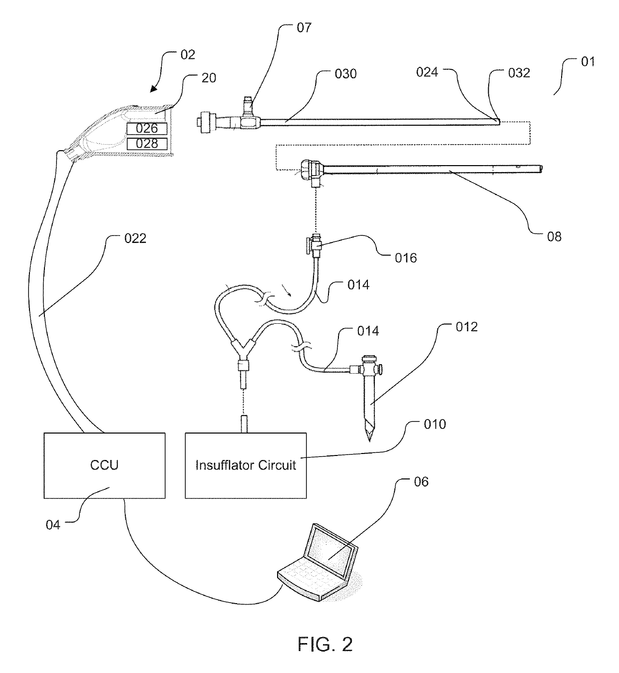
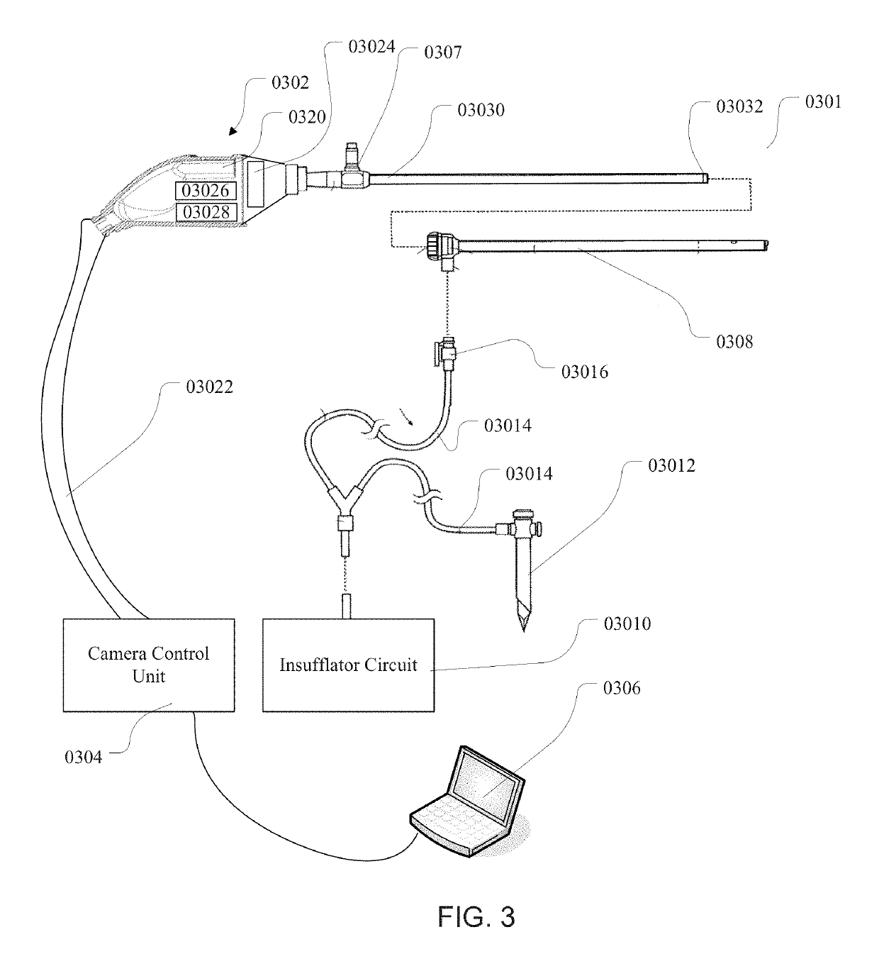
[0015]One aspect of the disclosure provides a view optimizing assembly having a deflector assembly with critical physical, pneumatic, and optical characteristics that make possible intra-operative defogging, surgical debris deflection, and cleaning of a laparoscope lens during minimally invasive surgery, while also maintaining visualization of the surgical site. In use, the view optimizing assembly makes possible the practice of a surgical method for maintaining clear visualization of the surgical site without removing the laparoscope 12 from the abdominal cavity for the purpose of cleaning or de-fogging its lens.
[0016]Another aspect of the disclosure provides a view optimizing assembly having a quick exchange feature. In use, the quick exchange feature makes possible a surgical method for maintaining clear visualization that includes the ability to make a quick exchange of laparoscopes having different operating characteristics (e.g., laparoscopes with different tip angles, lengths, or diameters) entirely on the sterile operating field and without interference with the preexisting surgical set-up on the sterile operating field. The view optimizing assembly integrates with the existing suite of minimally invasive instrumentation. It does not interfere with the surgical set-up, and it requires minimal change in the process or practice of a surgical operating room (OR) team.
[0020]In an embodiment an active imaging device may be attached to a control unit. The control unit will check the last sterilization date and ensure that the imaging device is no older than a predetermined safety date. If the imaging device is older than the required date, an onscreen warning will tell the user that the imaging device has expired and is unsafe for use. These features will protect the patient and the doctor from using a non-sterile imaging device.

Problems solved by technology

Due to this trend, it has become increasingly difficult to ensure that each endoscope and its components are properly cared for, used and sterilized for single use and not simply re-sterilized using traditional sterilization procedures.
Traditional drawbacks or problems of video endoscopes include a lack of image quality, the need for sterilization and high manufacturing cost as well as high processing cost.
The local environment within a patient's abdominal space is generally rather warm and humid, and the use of devices such as harmonic scalpels and other cutting and coagulating devices generate mist, smoke, and other debris that is released into the surgical field and often becomes suspended throughout the expanded abdominal space.
The need to remove the scope to defog and remove debris from the lens is inconvenient for the scope operator and the surgeon and can interrupt and undesirably prolong surgical procedures.

Method used


Image

  • Imaging sensor providing improved visualization for surgical scopes
  • Imaging sensor providing improved visualization for surgical scopes
  • Imaging sensor providing improved visualization for surgical scopes

Examples


Embodiment Construction

[0049]For the purposes of promoting an understanding of the principles in accordance with the disclosure, reference will now be made to the embodiments illustrated in the drawings and specific language will be used to describe the same. It will nevertheless be understood that no limitation of the scope of the disclosure is thereby intended. Any alterations and further modifications of the inventive features illustrated herein, and any additional applications of the principles of the disclosure as illustrated herein, which would normally occur to one skilled in the relevant art and having possession of this disclosure, are to be considered within the scope of the disclosure claimed.

[0050]Before the devices, systems, methods and processes for providing single use imaging devices and an image or view optimizing assembly are disclosed and described, it is to be understood that this disclosure is not limited to the particular embodiments, configurations, or process steps disclosed herein...


PUM


Abstract

A system, apparatus and methods for providing a single use imaging device having improved viewing for sterile environments is disclosed and described. A single use high definition camera used for general purpose surgical procedures including, but not limited to: arthroscopic, laparoscopic, gynecologic, and urologic procedures, may comprise an imaging device that is a sterile and designed to ensure single use. The imaging device may further include a sensor located near the tip of a laparoscope and a view optimizing assembly.

Description

CROSS-REFERENCE TO RELATED APPLICATIONS[0001]This application claims the benefit of U.S. Provisional Application No. 61 / 446,481, filed Feb. 24, 2011, which is hereby incorporated by reference herein in its entirety, including but not limited to those portions that specifically appear hereinafter, the incorporation by reference being made with the following exception: In the event that any portion of the above-referenced provisional application is inconsistent with this application, this application supercedes said above-referenced provisional application.STATEMENT REGARDING FEDERALLY SPONSORED RESEARCH OR DEVELOPMENT[0002]Not Applicable.BACKGROUND[0003]Endoscopic surgery is experiencing rapid growth in the medical field. Endoscopy is a minimally invasive surgical procedure that is used to analyze the interior of a body cavity or interior surfaces of an organ by inserting a tubular member into the body cavity through a minor or minimal incision. A conventional endoscope is generally ...

Claims


Application Information

Patent Timeline
Patent Type & Authority Patents(United States)
IPC IPC(8): A61B1/00A61B1/313A61B1/015A61B1/05
CPCA61B1/3132A61B1/00006A61B1/00009A61B1/015A61B1/05
Inventor TALBERT, JOSHUA D.HENLEY, JEREMIAH D.WICHERN, DONALD M.
Owner DEPUY SYNTHES PROD INC
Learn More