Cyclic RGD cell-binding motif and uses thereof
a cyclic rgd and cell-binding technology, applied in the field of new proteins, can solve the problems of affecting the effect of such experiments, affecting the and affecting the rational design of ligands for certain integrins, etc., and achieves the effect of promoting proliferation, differentiation and migration of cells
- Summary
- Abstract
- Description
- Claims
- Application Information
AI Technical Summary
Benefits of technology
Problems solved by technology
Method used
Image
Examples
example 1
ncorporation of Fibronectin-Derived Cell-Binding Motifs into Recombinant Spider Silk
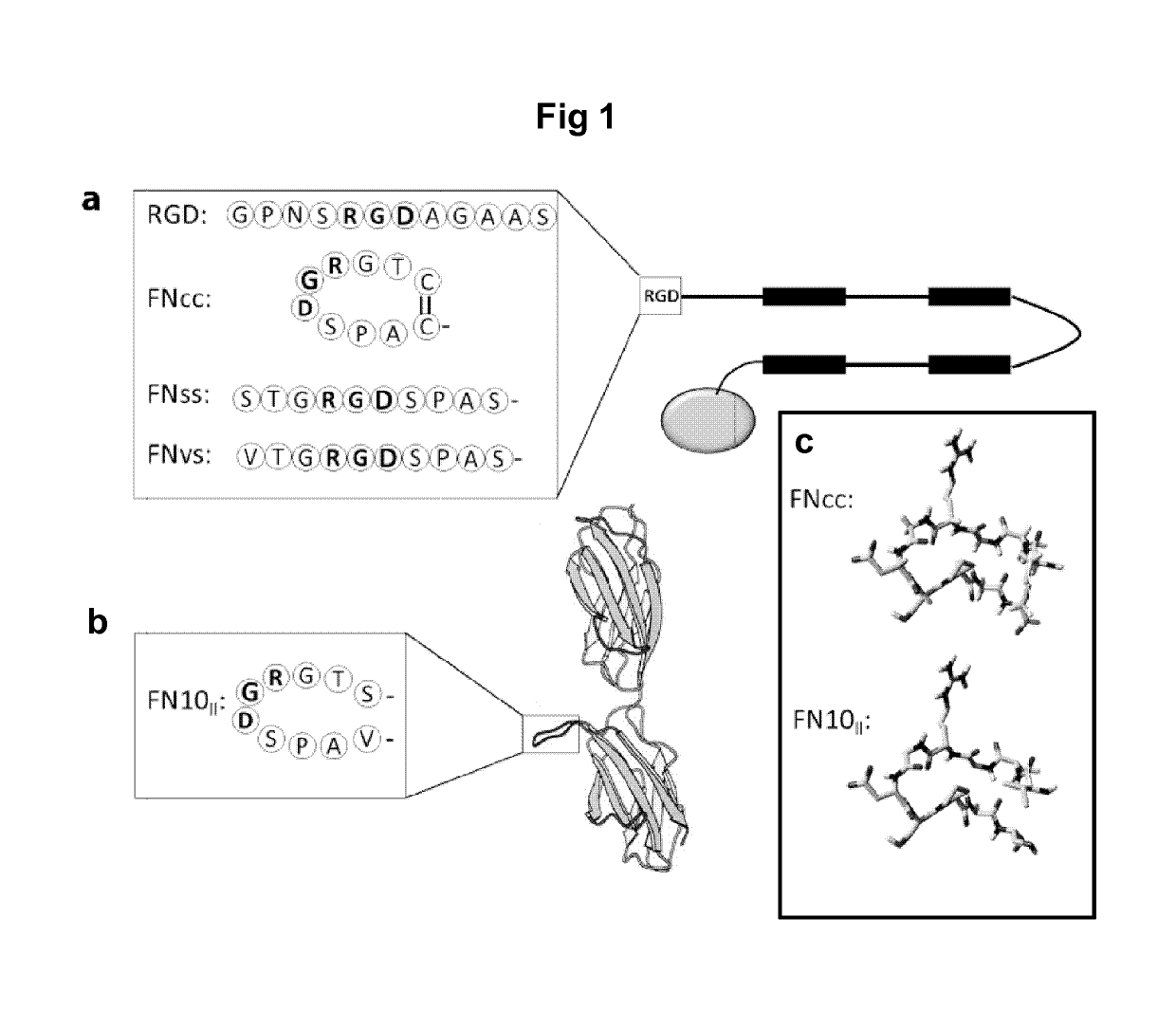
[0172]The recombinant spider silk protein 4RepCT (SEQ ID NO: 2, herein denoted WT) was genetically functionalized with the RGD containing cell binding motif from the fibronectin type III module 10, in four slightly different versions (FIG. 1). In the first (FNCC-4RepCT; SEQ ID NO: 13), two amino acids flanking the RGD sequence were substituted for cysteines to enable loop formation of the motif (CTGRGDSPAC; SEQ ID NO: 10). In the second (FNSS-4RepCT; SEQ ID NO: 14), the introduced cysteines were substituted for serines to create a linear control (STGRGDSPAS; SEQ ID NO: 11). Here the amino acid serine was selected due to its resemblance to cysteine, while lacking the ability to form disulfide bonds. In the third (FNVS-4RepCT; SEQ ID NO: 15), the original sequence of the fibronectin motif (VTGRGDSPAS; SEQ ID NO: 9) was used as a linear, native control. In the fourth (RGD-4RepCT; SEQ ID NO: 16), the RGD...
example 2
n of Fusion Proteins Containing Fibronectin-Derived Cell-binding Motifs
[0174]Protein production in E. coli of the genetic constructs obtained in Example 1 and the following purification were done essentially as described in Hedhammar M et al., Biochemistry 47(11):3407-3417 (2008) and Hedhammar M et al., Biomacromolecules 11: 953-959 (2010).
[0175]Briefly, Escherichia coli BL21(DE3) cells (Merck Biosciences) with the expression vector for the target protein were grown at 30° C. in Luria-Bertani medium containing kanamycin to an OD600 of 0.8-1 and then induced with isopropyl β-D-thiogalactopyranoside and further incubated for at least 2 h. Thereafter, cells were harvested and resuspended in 20 mM Tris-HCl (pH 8.0) supplemented with lysozyme and DNase I. After complete lysis, the supernatants from centrifugation at 15,000 g were loaded onto a column packed with Ni Sepharose (GE Healthcare, Uppsala, Sweden). The column was washed extensively before elution of bound proteins with 300 mM i...
example 3
on of Cell Culture Matrices
[0178]After purification, the protein solutions obtained in Example 2 were filter sterilized (0.22 μm) and concentrated by centrifugal filtration (Amicon Ultra, Millipore) before preparation of films, as described in Widhe M et al., Biomaterials 31(36): 9575-9585 (2010) and Widhe M et al., Biomaterials 34(33): 8223-8234 (2013).
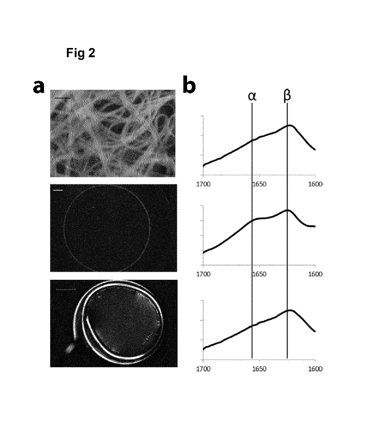
[0179]Briefly, petri dishes were coated at room temperature with recombinant spider silk solution at a concentration of 0.3 mg / ml to generate films. Foams were made by rapid pipetting of the silk solution, and fibers were formed by gentle wagging in 15 ml tube followed by cutting into smaller pieces.
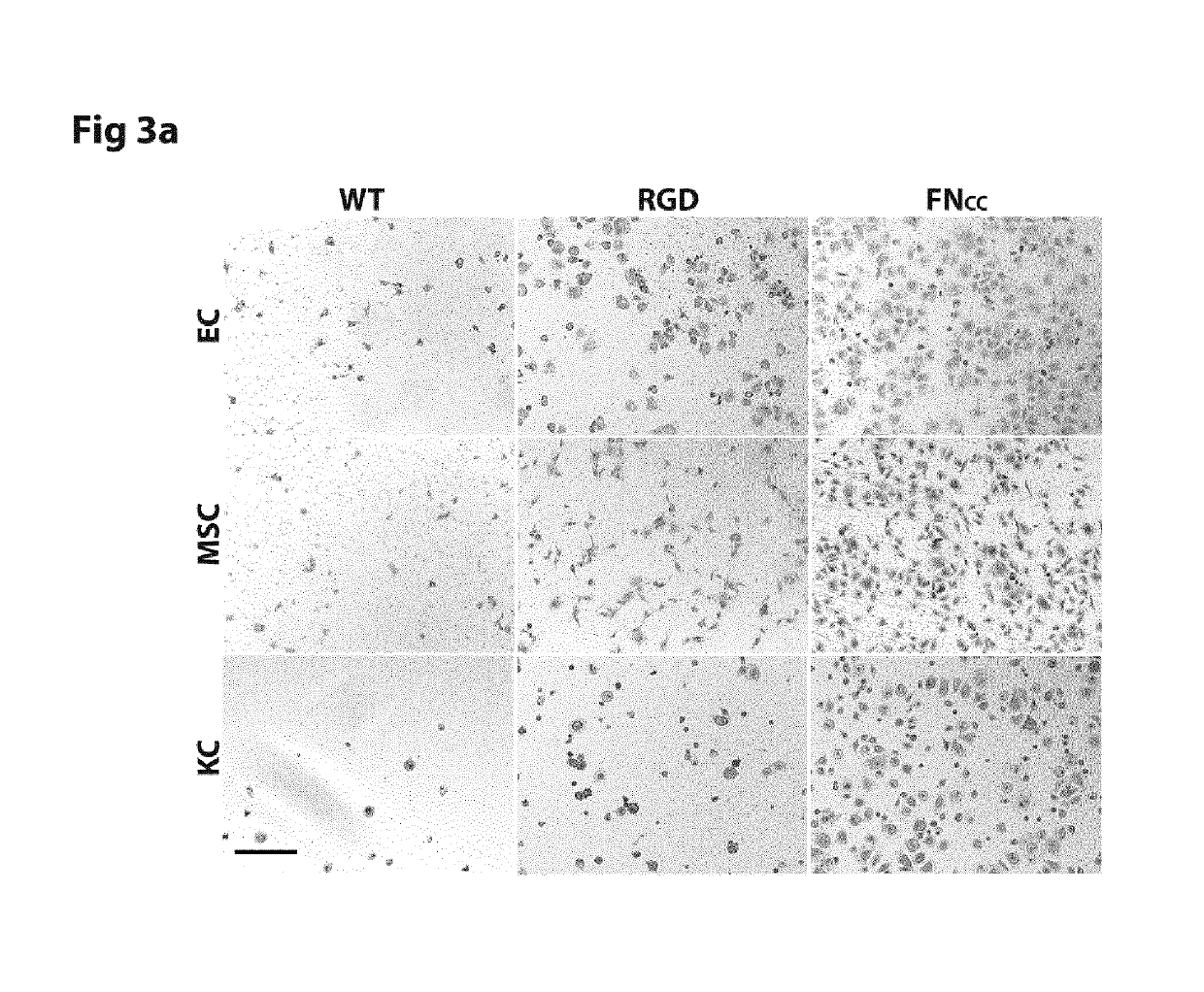
[0180]For studies of early attachment and repopulation, solutions of a protein concentration of 0.3 mg / ml were casted into films in 96- and 24 well cell culture plates respectively (Sarstedt, suspension cells) precoated with 1 pluronic to limit cell adhesion to the plastic surface. In control experiments, a reducing agent (either 5 mM Dit...
PUM
| Property | Measurement | Unit |
|---|---|---|
| size | aaaaa | aaaaa |
| thickness | aaaaa | aaaaa |
| thickness | aaaaa | aaaaa |
Abstract
Description
Claims
Application Information
 - R&D
- Intellectual Property
- Life Sciences
- Materials
- Tech Scout
- Unparalleled Data Quality
- Higher Quality Content
- 60% Fewer Hallucinations
Browse by: Latest US Patents, China's latest patents, Technical Efficacy Thesaurus, Application Domain, Technology Topic, Popular Technical Reports.
© 2025 PatSnap. All rights reserved.Legal|Privacy policy|Modern Slavery Act Transparency Statement|Sitemap|About US| Contact US: help@patsnap.com


