
A suprachoroidal space pressurized hydrogel balloon device for rhegmatogenous retinal detachment

A technique for retinal detachment and choroid, applied in the field of suprachoroidal space pressurized hydrogel balloon device, can solve the problems of poor controllability of final shape, inability to make effective adjustments, large difference in absorption time, etc., so as to reduce surgical pain and increase Surgical success rate, effect with small inter-individual differences

Inactive Publication Date: 2021-07-09
THE SECOND XIANGYA HOSPITAL OF CENT SOUTH UNIV
View PDF13 Cites 0 Cited by
  • Summary
  • Abstract
  • Description
  • Claims
  • Application Information

AI Technical Summary

Problems solved by technology

The controllability of this separation process and final shape is poor, and once the position of the pressurized crest deviates, it will not be able to make effective adjustments
Long-acting sodium hyaluronate is currently expensive, and the absorption time is relatively short, and the absorption time varies greatly among individuals. Therefore, suprachoroidal space compression needs to be further improved to improve its controllability and stability

Method used


Image

  • A suprachoroidal space pressurized hydrogel balloon device for rhegmatogenous retinal detachment
  • A suprachoroidal space pressurized hydrogel balloon device for rhegmatogenous retinal detachment
  • A suprachoroidal space pressurized hydrogel balloon device for rhegmatogenous retinal detachment

Examples


Embodiment 1

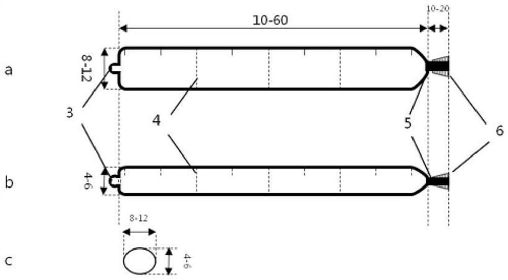
[0027] Such as figure 2 and Figure 4As shown, the present invention is composed of a hydrogel balloon 1, an injection needle 8 and a filler 2. The hydrogel balloon 1 is composed of a balloon main body, a head side blind end sheath 3, a tail end self-closing valve 5 and peripheral elastic bundles. The ring 6 is formed. When it is not inflated, the overall shape is a unilateral blind tube-like oblate sac. The tube wall of the cephalad blind-end sheath 3 is thicker than the wall of the main body of the balloon, forming a tip sheath-like structure that tightly wraps the injection needle 8. During the process of suprachoroidal space separation, the capsule wall and the surrounding tissue of the suprachoroidal space are effectively protected; after the filler 2 is injected, the sheath head of the blind end sheath 3 on the cephalic side expands with the hydrogel balloon 1, and is naturally unfolded and loosened for injection. The package of the needle 8; there is a one-way self-cl...


PUM


Abstract

The invention discloses a suprachoroidal space pressurized hydrogel balloon device for rhegmatogenous retinal detachment, which consists of a hydrogel balloon, an injection needle and a filler. The hydrogel balloon consists of a balloon main body, The head-side blind-end sheath and the tail-end self-closing valve are formed. When not inflated, the shape is a unilateral blind-tube-like oblate sac. The wall of the blind-end sheath on the head side of the balloon is thicker than the wall of the main body of the balloon, forming a tight wrap around the tip of the injection needle. There is a one-way self-closing diaphragm in the main body of the balloon, and the long diameter of the balloon is divided into multiple compartments. The tail side of the balloon is connected with a hydrogel double self-closing valve, and the inner diameter of the tube can be placed in the suprachoroidal space After the needle core is punctured and the injection needle is withdrawn, the lumen is tightly closed due to the dual effects of the elastic retraction of the tube wall itself and the corresponding peripheral elastic ring, so that the contents of the balloon will not leak out; there is a scale on the injection needle. The length, width, and height of the balloon pressurized crest of the present invention are stable, controllable and adjustable, the absorption time of the balloon wall determines the retention time of the crest, and the difference among individuals is small, which can improve the success rate of the operation.

Description

technical field [0001] The invention relates to a surgical instrument used in ophthalmic retinal diseases in clinical medicine, in particular to a suprachoroidal space pressurized hydrogel balloon device for rhegmatogenous retinal detachment. Background technique [0002] Rhegmatogenous retinal detachment is the most common blinding retinal disease, which is caused by the separation of retinal neuroepithelium and pigment epithelium due to a tear in the retinal neuroepithelium and fluid in the vitreous entering the subretina. There are more males than females at about 3:2, and most of them are adults over 30 years old. There is a difference between the left and right eyes, and the incidence of both eyes is about 15% of the total number of patients. Occur in myopia, especially high myopia. [0003] The basic principle of treatment of rhegmatogenous retinal detachment is to seal the hole, promote the absorption of subretinal fluid and calm the retina, which can be roughly divi...

Claims


Application Information

Patent Timeline
Patent Type & Authority Patents(China)
IPC IPC(8): A61F9/007
CPCA61F9/0008A61F9/00727A61F9/00736
Inventor 李芸赵元曹健陈百华谭周辉
Owner THE SECOND XIANGYA HOSPITAL OF CENT SOUTH UNIV
Learn More